. 2006 Mar 20;203(3):541-52.
doi: 10.1084/jem.20050407. Epub 2006 Feb 27.

The primary defect in experimental ileitis originates from a nonhematopoietic source

Affiliations

The primary defect in experimental ileitis originates from a nonhematopoietic source

Timothy S Olson et al. J Exp Med. .

Abstract

The initiating etiologic factor in Crohn's disease (CD) remains unclear. SAMP1/YitFc (SAMP) mice develop chronic ileitis similar to human CD. We used bone marrow chimeras to determine if SAMP ileitis results from a primary immunological defect or from dysregulated mucosal immunity secondary to intrinsic, nonhematopoietic (e.g., epithelial) dysfunction. SAMP mice receiving wild-type (AKR) BM developed severe ileitis, whereas SAMP BM did not confer ileitis to WT recipients. WT lymphocytes from reconstituted SAMP mice resembled native SAMP populations in regard to surface phenotype and cytokine production. Ilea from native SAMP mice and SAMP recipients of wild-type BM displayed decreased epithelial barrier resistance ex vivo and increased epithelial permeability in vivo compared to native WT mice and AKR recipients of SAMP BM. This permeability defect preceded the development of ileal inflammation, was present in the absence of commensal bacteria, and was accompanied by altered ileal mRNA expression of the tight junction proteins claudin-2 and occludin. Our results provide evidence that the primary defect conferring ileitis in SAMP mice originates from a nonhematopoietic source. Generation of pathogenic lymphocytes is a consequence of this defect and does not reflect intrinsic proinflammatory leukocyte properties. Decreased barrier function suggests that defects in the epithelium may represent the primary source of SAMP ileitis susceptibility.


Figures

Figure 1.
Figure 1.
Ileitis is dependent on host/nonhematopoietic compartment and not donor BM. (A) Disease severity was assessed in SAMP (n = 7), AKR (n = 5), SAMP BM → SAMP (n = 5), AKR BM → SAMP (n = 10), AKR BM → AKR (n = 3), and SAMP BM → AKR (n = 10) mice. *Significant difference in inflammatory score between SAMP BM → AKR and AKR BM → SAMP mice. (B) Histologic evaluation of ileal sections from SAMP and AKR BM → SAMP mice shows large inflammatory infiltrates and epithelial abnormalities including crypt hypertrophy and elongation (arrowheads), and Paneth cell hyperplasia (arrows). Sections from AKR and SAMP BM → AKR mice display normal architecture.
Figure 2.
Figure 2.
Inflammatory status of host determines pathogenicity and phenotype of MLN lymphocytes. Flow cytometry was performed on MLN lymphocyte populations in SAMP (n = 3), AKR (n = 3), AKR BM → SAMP (n = 7), and SAMP BM → AKR (n = 6) mice. (A) Representative T cell–gated (CD4+CD8+) dotplots of CD4 (y) versus CD69 (x) expression, with percentages of CD4+ T cells that are CD69+. (B) Representative dotplots of B220 (x) versus IgA (y) expression, with averages of IgA+B220hi mature B cells and IgA+B220int plasmablasts as a percentage of all lymphocytes. Results are expressed as mean ± SEM. *Indicates significantly increased (P < 0.05) compared with AKR or SAMP BM → AKR mice. Lymphocyte subsets that were significantly increased in SAMP (x) versus AKR (y) mice included the percentage of IgA+ cells (♦) and the percentage of CD4+ T cells that were αE + (▵), β7 hi (□), CD25+ (•), CD44hi (▴), CD69+ (⋄), and l-selectinlo (▪) (C, left). Similar trends were seen in a plot of SAMP BM → AKR (y) versus AKR BM → SAMP (x) subsets (C, right). The slopes of the best-fit lines through the points in the two plots were not significantly different.
Figure 3.
Figure 3.
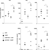
Increased cytokine expression in MLN CD4+ T cells from AKR BM → SAMP mice. Levels of secreted TNF, IFNγ, IL-2, IL-4, and IL-5 were measured in triplicate by cytometric bead array, from 48-h cultures of AKR (□, n = 3), SAMP (▪, n = 4), SAMP BM → AKR (▵, n = 3), and AKR BM → SAMP (▴, n = 4) MLN CD4+ T cells (105/well), stimulated with immobilized anti-CD3 antibody. Points represent individual mice with the mean value (_) in each group also shown. *Significantly increased compared with SAMP BM → AKR cells (P < 0.05).
Figure 4.
Figure 4.
Epithelial barrier dysfunction in SAMP and AKR BM → SAMP mice. (A) TEER assays on ileum and jejunum from SAMP (n = 8), AKR (n = 8), SAMP BM → SAMP (n = 5), AKR BM → SAMP (n = 4), AKR BM → AKR (n = 3), and SAMP BM → AKR (n = 4) mice. Ilea from SAMP, SAMP BM → SAMP, and AKR BM → SAMP mice exhibited decreases in ΔTEER indicative of increased epithelial permeability, whereas ilea from AKR, AKR BM → AKR, and SAMP BM → AKR mice maintained an effective epithelial resistance barrier. Significant differences (P < 0.03) are indicated between experimental groups (A, left). No differences were observed in jejunal TEER among BMT recipient groups. A significant in crease in TEER was seen in native SAMP compared with AKR jejunum; P < 0.03 (A, right). (B) In vivo assays measuring epithelial permeability to solutes in the small intestine and colon using urinary FE ratio of lactulose to mannitol (B, left) and of sucralose (B, right), respectively, in AKR BM → SAMP (♦) (n = 6) and SAMP BM → AKR (▪) (n = 6) mice. The time 0 point was collected immediately before irradiation and transplantation and thus represent the permeability of native SAMP (♦) and AKR (▪) tissues. *Indicates significantly increased (P < 0.05) compared with SAMP BM → AKR mice.
Figure 5.
Figure 5.
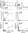
Increased small intestinal permeability occurs before the onset of inflammation in SAMP mice. (A) Paracellular permeability, measured in the small intestine and colon using the in vivo probe assay, was increased as early as 3 wk of age in SAMP (□) compared with age-matched AKR (▪) mice, and remained elevated through 20 wk. (B) Total inflammatory scores for the ileum and colon show that inflammation was not significantly increased in SAMP versus AKR ileum until 12 wk of age. (C) Ileal MPO activity, providing a quantitative marker of active inflammation, showed a strong correlation with histologic ileal inflammatory score (r = 0.77) and was also not increased in SAMP versus AKR mice until 12 wk. n ≥ 4 per group; *P < 0.05 versus age-matched AKR.
Figure 6.
Figure 6.
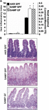
Early, preinflammatory alterations in small intestinal barrier function persist in the absence of the enteric flora. (A) Small intestinal paracellular permeability in GF SAMP mice was similar to permeability in SAMP mice raised under SPF conditions, and significantly increased compared with SPF-AKR permeability n ≥ 4 per group; *P < 0.05 versus age-matched AKR. (B) 3-wk-old GF SAMP mice displayed no histological signs of ileal inflammation and possessed normal intestinal architecture.
Figure 7.
Figure 7.
Altered epithelial TJ protein expression in young, uninflamed SAMP compared with AKR mice. mRNA transcript levels of TJ proteins (claudin [CLDN] 1–4 and occludin) were measured by RT-PCR and expressed relative to HGPRT in isolated intestinal epithelial cell preparations from the ileum (left) or colon (right) of SAMP and AKR mice (3 wk of age; n ≥ 4 animals per group). The greatest differences in expression in SAMP ileum compared with age-matched AKR controls were seen in CLDN2 (eightfold increase; top left) and occludin (1.8-fold decrease; bottom left). Similar trends were seen in the colon, though increases in CLDN2 (top right) expression in SAMP versus AKR colon were not statistical significant. No differences in expression were noted for CLDN1, 3, and 4 in either the ileum or colon of SAMP relative to controls. n ≥ 4 per group; *P < 0.05 versus control AKR.

References

    1. Bouma, G., and W. Strober. 2003. The immunological and genetic basis of inflammatory bowel disease. Nat. Rev. Immunol. 3:521–533. - PubMed
    1. Egan, L.J., and W.J. Sandborn. 2004. Advances in the treatment of Crohn's disease. Gastroenterology. 126:1574–1581. - PubMed
    1. Ma, T.Y. 1997. Intestinal epithelial barrier dysfunction in Crohn's disease. Proc. Soc. Exp. Biol. Med. 214:318–327. - PubMed
    1. Shanahan, F. 2002. Crohn's disease. Lancet. 359:62–69. - PubMed
    1. Ogura, Y., N. Inohara, A. Benito, F.F. Chen, S. Yamaoka, and G. Nunez. 2001. Nod2, a Nod1/Apaf-1 family member that is restricted to monocytes and activates NF-kappaB. J. Biol. Chem. 276:4812–4818. - PubMed

Publication types

MeSH terms