. 2006 Mar 20;203(3):661-73.
doi: 10.1084/jem.20051777. Epub 2006 Feb 27.

A structural basis for selection and cross-species reactivity of the semi-invariant NKT cell receptor in CD1d/glycolipid recognition

Affiliations

A structural basis for selection and cross-species reactivity of the semi-invariant NKT cell receptor in CD1d/glycolipid recognition

Lars Kjer-Nielsen et al. J Exp Med. .

Abstract

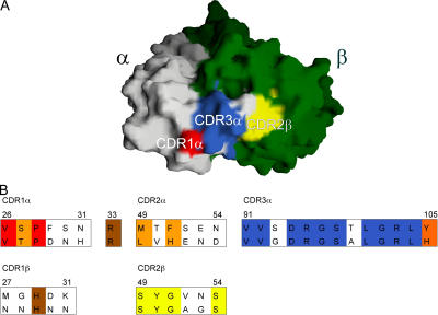
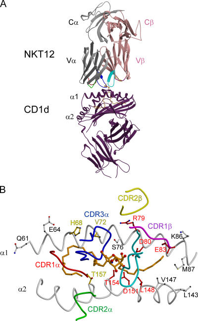
Little is known regarding the basis for selection of the semi-invariant alphabeta T cell receptor (TCR) expressed by natural killer T (NKT) cells or how this mediates recognition of CD1d-glycolipid complexes. We have determined the structures of two human NKT TCRs that differ in their CDR3beta composition and length. Both TCRs contain a conserved, positively charged pocket at the ligand interface that is lined by residues from the invariant TCR alpha- and semi-invariant beta-chains. The cavity is centrally located and ideally suited to interact with the exposed glycosyl head group of glycolipid antigens. Sequences common to mouse and human invariant NKT TCRs reveal a contiguous conserved "hot spot" that provides a basis for the reactivity of NKT cells across species. Structural and functional data suggest that the CDR3beta loop provides a plasticity mechanism that accommodates recognition of a variety of glycolipid antigens presented by CD1d. We propose a model of NKT TCR-CD1d-glycolipid interaction in which the invariant CDR3alpha loop is predicted to play a major role in determining the inherent bias toward CD1d. The findings define a structural basis for the selection of the semi-invariant alphabeta TCR and the unique antigen specificity of NKT cells.


Figures

Figure 1.
Figure 1.
Structural and functional integrity of recombinant soluble NKT TCRs. (A) Purified bacterial NKT15 TCR was analyzed by SDS-PAGE under reducing (+DTT, dithiothreitol) and nonreducing (−DTT) conditions demonstrating αβ heterodimers. (B) Native gel electrophoresis of folded αβ heterodimeric NKT15 and control LC13 TCRs. (C) Recombinant soluble NKT12, NKT15, NKT18, and control LC13 TCRs were tested for their ability to block binding of mouse CD1d/α-GalCer tetramers to murine thymocytes. Phycoerythrin-conjugated mCD1d/α-GalCer tetramers were preincubated with the indicated soluble TCRs over a range of TCR concentrations before staining mouse thymocytes. Cells were analyzed by two-color flow cytometry showing mCD1d/α-GalCer tetramer staining on the vertical axis and FITC-CD3 (mAb 145-2C11) staining on the horizontal axis. Cells staining positively with mCD1d/α-GalCer tetramer and FITC-CD3 are indicated with a circle.
Figure 2.
Figure 2.
Overview of NKT cell TCR structure showing the conformation of the CDR loops at the Ag-binding interface. (A) Overview of the structure of NKT12 with the α-chain and β-chain shown in gray and pink, respectively. (B) The CDR3 loops of NKT12 are color coded according to their genetic origin. (C) Superimposition of the CDR loops of the NKT12 and NKT15 TCRs depicting the difference in the conformation of CDR3β (NKT12, CDR loops colors are indicated; NKT15, orange).
Figure 3.
Figure 3.
The Ag-binding interface of NTK12 and NKT15 TCRs contains a preformed cavity created by invariant residues of the α-chain, a species-conserved residue from the β-chain and CDR3β. (A) NKT12 TCR α-chain residues that are conserved between human and mouse are red; α-chain residues that differ between these species are gray. The TCR β-chain residue Tyr33β is conserved across species, whereas the CDR3β residues are divergent between species. β-chain side chains are green. (B) Close-up of residues forming the putative ligand-binding cavity of the NKT12 binding surface. Residues are labeled according to the single amino acid code. (C) The putative ligand-binding cavity of NKT15 is obstructed by the bulky side chain of residue Tyr 103β from CDR3β in the trigonal form of the crystal structure. (D) Tyr103β could not be visualized in the NKT15 TCR crystal comprising the orthorhombic space group. The high mobility of Tyr103β could easily allow displacement of this side chain, exposing the putative Ag-binding cavity. Accordingly, the Tyr103β side chain has been omitted, revealing the cavity.
Figure 4.
Figure 4.
NKT cell receptor CDR3β regions impact on the recognition of CD1d/α-GalCer. (A) Conformation of the CDR3 loops of the NKT15 TCR revealing the highly exposed “RDR” motif at the tip of the CDR3β loop. (B) Inhibition of NKT cell staining by recombinant wild-type and chimeric NKT TCRs. TCRs comprised LC13αβ; NKT15αβ; NKT15α-chain/LC13 β-chain; NKT15α-chain/NKT15 β-chain engrafted with the LC13 CDR3β-loop; and NKT15α-chain/NKT15β-chain with “RDR” to “AAA” substitution within the CDR3β loop. Graded concentrations of soluble recombinant TCRs were preincubated with phycoerythrin-labeled tetramers of mCD1dα-GalCer. These were used to costain mouse thymocyte cells with anti-CD3 mAb and analyzed by flow cytometry. Shown are flow cytometry plots with tetramer-staining after preincubation with 500 μg/ml TCRs. *, the inhibition observed with maximal concentrations of the NKTα+LC13(CDR3β) (500 μg/ml) was only partial, and equivalent to the inhibition observed with 8 μg/ml (not depicted) of NKT15. The indicated KD values are calculated from SPR studies of TCR binding to immobilized human and mouse CD1dα-GalCer (Table S3 and Fig. S2 for NKT12 and NKT15). LC13 is a control TCR from an HLA-B8–restricted, virus-specific CTL; ND, KD not measurable. (C) Superposition of the CDR3β loop of LC13 in the context of the NKT CDR3α based on the known structure of LC13 (references 36, 37). The LC13 CDR3β loop is predicted to impact on the conformation of CDR3α and to disrupt access to the putative Ag-binding cavity.
Figure 5.
Figure 5.
Conserved residues from human and mouse NKT TCR CDR1α, CDR3α, and CDR2β loops form a contiguous conserved surface that is adjacent to the putative Ag-binding cavity. The different conserved CDR regions are colored to correspond to the sequence alignment of mouse and human CDR loops. All other conserved and semi-conserved residues are brown and orange, respectively, in the alignment and are not shown in the structure. The TcR α-chain is white and the TCR β-chain is green.
Figure 6.
Figure 6.
Proposed docking of NKT TCR onto CD1d/α-GalCer. (A) In the model, the NKT12 α and β chain are shown in gray and pink, respectively, and the CDR loops are red (CDR1α), green (CDR2α), blue (CDR3α), magenta (CDR1β), yellow (CDR2β), and cyan (CDR3β). α-galactosylceramide is shown in orange within the antigen-binding domain of hCD1d (purple). (B) View of the antigen-binding cleft of human CD1d–presenting α-GalCer (orange). The CDR footprint of the proposed docking model of NKT12 is shown for reference and the CDR loops are colored as in A. Surface-exposed residues on the α1 and α2 helices of hCD1d are shown. Residues in red had an impact on NKT activation by mCD1d in the published literature. Residues in white either had no impact or were not investigated. Residues in yellow are those residues likely to be important in the docking of NKT12 onto CD1d/α-galactosylceramide according to our proposed model.

References

    1. Ulrichs, T., and S.A. Porcelli. 2000. CD1 proteins: targets of T cell recognition in innate and adaptive immunity. Rev. Immunogenet. 2:416–432. - PubMed
    1. Brigl, M., and M.B. Brenner. 2004. CD1: antigen presentation and T cell function. Annu. Rev. Immunol. 22:817–890. - PubMed
    1. Kobayashi, E., K. Motoki, T. Uchida, H. Fukushima, and Y. Koezuka. 1995. Krn7000, a novel immunomodulator, and its antitumor activities. Oncol. Res. 7:529–534. - PubMed
    1. Hansen, D.S., M.A. Siomos, L. Buckingham, A.A. Scalzo, and L. Schofield. 2003. Regulation of murine cerebral malaria pathogenesis by CD1d-restricted NKT cells and the natural killer complex. Immunity. 18:391–402. - PubMed
    1. Schofield, L., M.J. McConville, D. Hansen, A.S. Campbell, B. Fraser-Reid, M.J. Grusby, and S.D. Tachado. 1999. CD1d-restricted immunoglobulin G formation to GPI-anchored antigens mediated by NKT cells. Science. 283:225–229. - PubMed

Publication types

MeSH terms