. 2006 Feb 27;172(5):705-17.
doi: 10.1083/jcb.200508166.

Recycling of ESCRTs by the AAA-ATPase Vps4 is regulated by a conserved VSL region in Vta1

Affiliations

Recycling of ESCRTs by the AAA-ATPase Vps4 is regulated by a conserved VSL region in Vta1

Ishara Azmi et al. J Cell Biol. .

Abstract

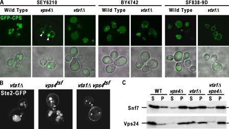
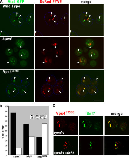
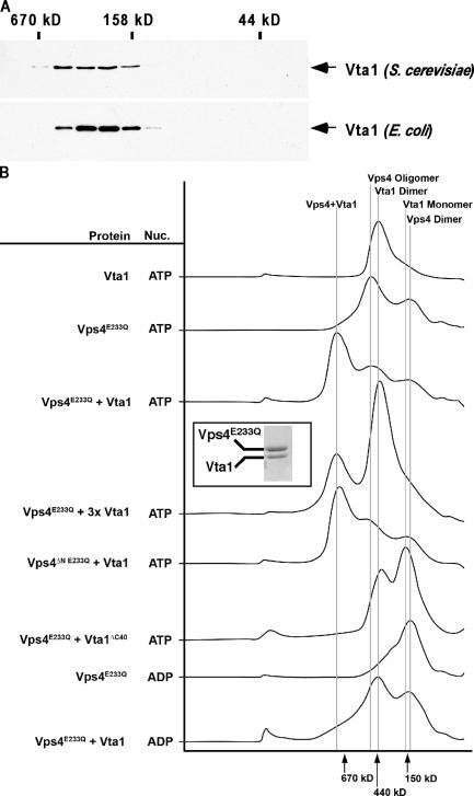
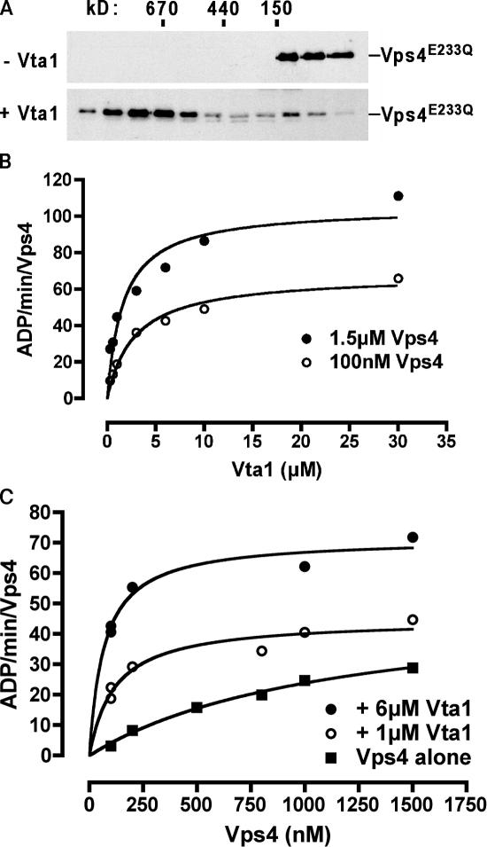
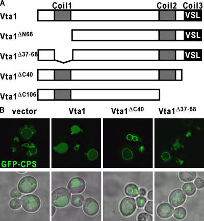
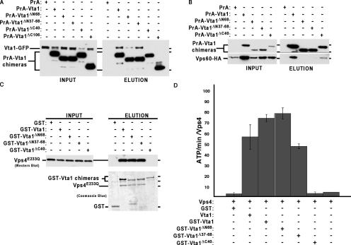
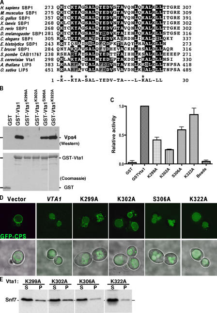
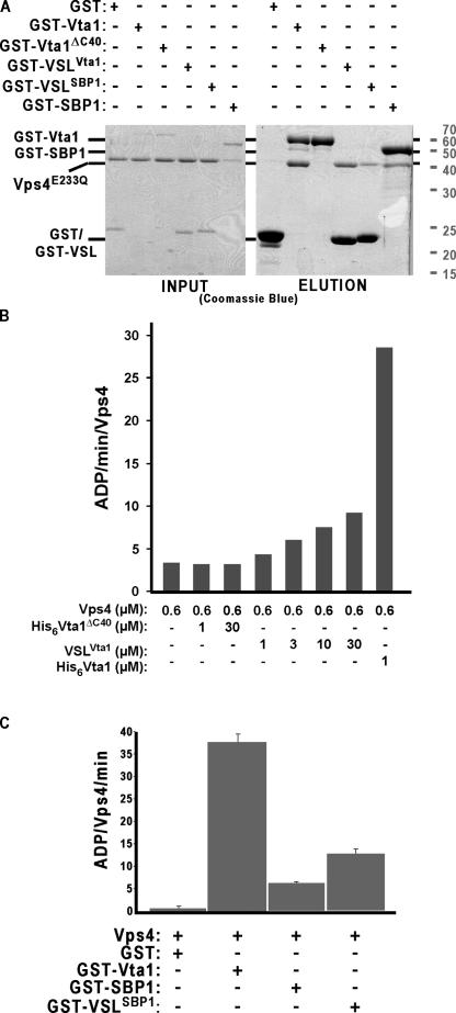
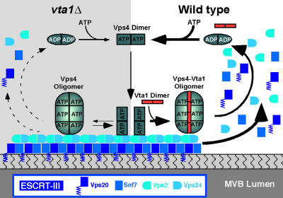
In eukaryotes, the multivesicular body (MVB) sorting pathway plays an essential role in regulating cell surface protein composition, thereby impacting numerous cellular functions. Vps4, an ATPase associated with a variety of cellular activities, is required late in the MVB sorting reaction to dissociate the endosomal sorting complex required for transport (ESCRT), a requisite for proper function of this pathway. However, regulation of Vps4 function is not understood. We characterize Vta1 as a positive regulator of Vps4 both in vivo and in vitro. Vta1 promotes proper assembly of Vps4 and stimulates its ATPase activity through the conserved Vta1/SBP1/LIP5 region present in Vta1 homologues across evolution, including human SBP1 and Arabidopsis thaliana LIP5. These results suggest an evolutionarily conserved mechanism through which the disassembly of the ESCRT proteins, and thereby MVB sorting, is regulated by the Vta1/SBP1/LIP5 proteins.


Figures

Figure 1.
Figure 1.
MVB sorting defect in vta1Δ is distinct from the known class E vps mutant. (A) The MVB cargo GFP-CPS was visualized by fluorescent and brightfield microscopy in living cells corresponding to wild type, vta1Δ, or the known class E vps mutant vps4Δ in three distinct genetic backgrounds. Arrowheads highlight the class E compartment in vps4Δ cells. (B) Synthetic genetic interaction between vta1Δ and vps4tsf. Ste2-GFP was visualized by fluorescent microscopy in vta1Δ, vps4tsf, and vta1Δ vps4tsf genetic backgrounds at permissive temperature. (C) Vta1 is required for efficient release of the ESCRT-III subunits Snf7 and Vps24 from endosomal membranes. Differential centrifugation was performed on wild-type, vps4Δ, vta1Δ, and vps4Δ vta1Δ cell extracts. The resulting soluble (S) and pellet (P) fractions were probed with anti-Snf7 or anti-Vps24 antibodies.
Figure 2.
Figure 2.
Interactions of Vta1 and Vps4 in vivo. (A) Vta1 requires Vps4 for endosomal association. Fluorescence microscopy was performed on wild-type, vps4Δ, and vps4E233Q cells expressing Vta1-GFP and the endosomal marker DsRed-FYVE. Arrowheads indicate endosomes in wild-type cells or class E compartments in vps4Δ and vps4E233Q. Dashed blue lines are used to outline cells. Bar, 5 μM. (B) Quantification of endosomal association of Vta1 in wild type, vps4Δ, and vps4E233Q. Differential centrifugation was performed on extracts made from wild-type, vps4Δ, and vps4E233Q cells expressing Vta1-HAand probed with anti-HA antibody. Immunoreactive species were quantitated and graphed as the percentage present in soluble and membrane fractions. This experiment was performed twice, and representative data is shown from one of the experiments. (C) Vta1 is not required for endosomal localization of Vps4. Immunofluorescence was performed to visualize the subcellular localization of Vps4 and Snf7 in either vps4Δ or vps4Δ vta1Δ cells expressing Vps4E233Q-HA. Bar, 5 μm.
Figure 3.
Figure 3.
Vta1 homooligomerization and heterooligomerization with Vps4. (A) Vta1 exists as a homooligomer in solution. Sephacryl S-200 gel filtration analyses were performed on yeast extracts or bacterial extracts, and Vta1-HA or His6-Vta1 was visualized with appropriate antibodies. (B) Vta1 and Vps4 form a higher order protein complex in the presence of ATP. Purified recombinant Vta1 and Vps4E233Q were subjected to Superose 6 gel filtration analysis under a variety of conditions. UV traces from the fast protein liquid chromatography runs, wherein the y axis corresponds to absorbance at 280 nm and the x axis corresponds to the elution volume, are presented to summarize results. The peak of the 1-MD complex was subjected to SDS-PAGE and Coomassie blue staining, displayed in the inset.
Figure 4.
Figure 4.
Vta1 stimulates Vps4 ATPase activity. (A) Vta1 stimulates Vps4 assembly into its ATPase-competent form. A low concentration of Vps4 was subjected to Superose 6 gel filtration analyses (containing ATP as in Fig. 3 B) in the presence or absence of Vta1, and immunoreactive species were detected by Western blotting with anti-Vps4 antisera. (B) ATPase activity of Vps4 in the presence of Vta1, measured as a function of Vta1 concentration. (C) ATPase activity of Vps4 in the presence of Vta1, measured as a function of Vps4 concentration. Values in both B and C are presented as specific activity per Vps4 molecule. Prism 4 software was used to fit data points to a curve with nonlinear regression.
Figure 5.
Figure 5.
Coiled-coil domains of Vta1 are required for function. (A) Schematic representation of Vta1 and mutant forms. Putative coiled-coil domains are indicated, as is the VSL region. (B) In vivo analysis of Vta1 coiled-coil mutants. BY4742 cells deleted for VTA1 were transformed with PrA-VTA1 chimeras as indicated, and MVB sorting was addressed by visualization of GFP-CPS.
Figure 6.
Figure 6.
Domain analysis of Vta1. (A) Cells expressing Vta1-GFP were transformed with plasmids expressing PrA, PrA-Vta1, or mutant versions thereof; native lysates were generated; PrA fusion proteins were isolated using IgG Sepharose; and material was analyzed by Western blotting with anti-GFP antibody. (B) In vivo analysis of Vta1–Vps60 interactions. Cells were transformed with a plasmid expressing an HA-tagged form of Vps60, along with PrA-Vta1 constructs; native lysates were generated; PrA fusions were isolated using IgG Sepharose; and material was analyzed by Western blotting with anti-HA antibody. (C) In vitro analysis of Vta1–Vps4 interactions. Purified Vps4E233Q was incubated with GST or GST fusion proteins corresponding to Vta1 or mutant forms thereof; glutathione Sepharose was used as an affinity isolation step; and material was visualized by Western blotting with Vps4 or Coomassie blue staining. (D) ATPase activity of 0.6 μM Vps4 in the presence of mutated forms of Vta1. n = 3.
Figure 7.
Figure 7.
An evolutionarily conserved motif in the COOH terminus of Vta1 homologues is necessary for Vps4 binding and stimulation of ATPase activity. (A) Alignment of the VSL region from Vta1 and homologues. Amino acids that have been altered are indicated by asterisks. (B) Vps4 binding by VSL point mutants. Purified Vps4E233Q was incubated with purified GST, GST-Vta1, or the indicated point mutant thereof; glutathione Sepharose was used as an affinity isolation step; and bound material was analyzed by Coomassie staining or anti-Vps4 Western blotting. (C) Stimulation of Vps4 ATPase activity by VSL point mutants. Vps4 ATPase activity was measured in the conditions indicated and normalized to wild-type Vta1 to calculate relative stimulation. n = 3. Error bars indicate SEM. (D) Effect of VSL point mutations on MVB sorting reaction. GFP-CPS was visualized in vta1Δ cells (BY4742) expressing the indicated form of Vta1. (E) Mutations in the VSL region perturb ESCRT-III recycling. Differential centrifugation was performed on vta1Δ cells expressing the indicated form of Vta1. The resulting soluble (S) and pellet (P) fractions were probed with anti-Snf7 antibody. White lines indicate that intervening lanes have been spliced out.
Figure 8.
Figure 8.
The VSL domain of Vta1 and SBP1 is sufficient for Vps4 binding. (A) Purified Vps4E233Q was incubated with purified GST, GST-Vta1, GST-Vta1ΔC40, GST-VSLVta1, GST-SBP1, or GST-VSLSBP1; glutathione Sepharose was used as an affinity isolation step; and material was visualized by staining with Coomassie blue. (B) ATPase activity of Vps4 is stimulated by the VSL domain of Vta1. This experiment was performed twice, and representative data is presented from one of those experiments. (C) ATPase activity of Vps4 is stimulated by hSBP1 and its VSL domain. n = 4. Error bars indicate SEM.
Figure 9.
Figure 9.
Model for Vta1–Vps4 function in MVB sorting. ATPase activity of Vps4 requires the assembly of ATP-bound Vps4 dimers into an oligomeric form. The ATPase function of Vps4 is required for proper function of the MVB sorting pathway. In wild-type cells, assembly of oligomeric Vps4 and associated ATPase activity is stimulated by Vta1. This Vta1–Vps4 oligomer associates with the endosomal membrane via ESCRT-III subunits to potentiate the dissociation of ESCRTs. In vta1Δ cells, Vps4 function is partially compromised and ESCRT membrane association is partially stabilized.

References

    1. Amerik, A.Y., J. Nowak, S. Swaminathan, and M. Hochstrasser. 2000. The Doa4 deubiquitinating enzyme is functionally linked to the vacuolar protein-sorting and endocytic pathways. Mol. Biol. Cell. 11:3365–3380. - PMC - PubMed
    1. Babst, M. 2005. A protein's final ESCRT. Traffic. 6:2–9. - PubMed
    1. Babst, M., T.K. Sato, L.M. Banta, and S.D. Emr. 1997. Endosomal transport function in yeast requires a novel AAA-type ATPase, Vps4p. EMBO J. 16:1820–1831. - PMC - PubMed
    1. Babst, M., B. Wendland, E.J. Estepa, and S.D. Emr. 1998. The Vps4p AAA ATPase regulates membrane association of a Vps protein complex required for normal endosome function. EMBO J. 17:2982–2993. - PMC - PubMed
    1. Babst, M., D.J. Katzmann, E.J. Estepa-Sabal, T. Meerloo, and S.D. Emr. 2002. a. Escrt-III: an endosome-associated heterooligomeric protein complex required for mvb sorting. Dev. Cell. 3:271–282. - PubMed

Publication types

MeSH terms

Substances