. 2006 Feb 27;172(5):733-45.
doi: 10.1083/jcb.200511068.

A motor neuron disease-associated mutation in p150Glued perturbs dynactin function and induces protein aggregation

Affiliations

A motor neuron disease-associated mutation in p150Glued perturbs dynactin function and induces protein aggregation

Jennifer R Levy et al. J Cell Biol. .

Abstract

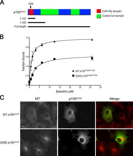
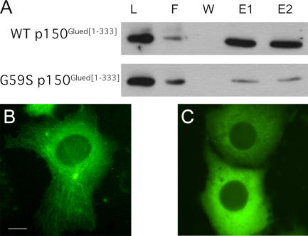
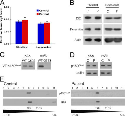
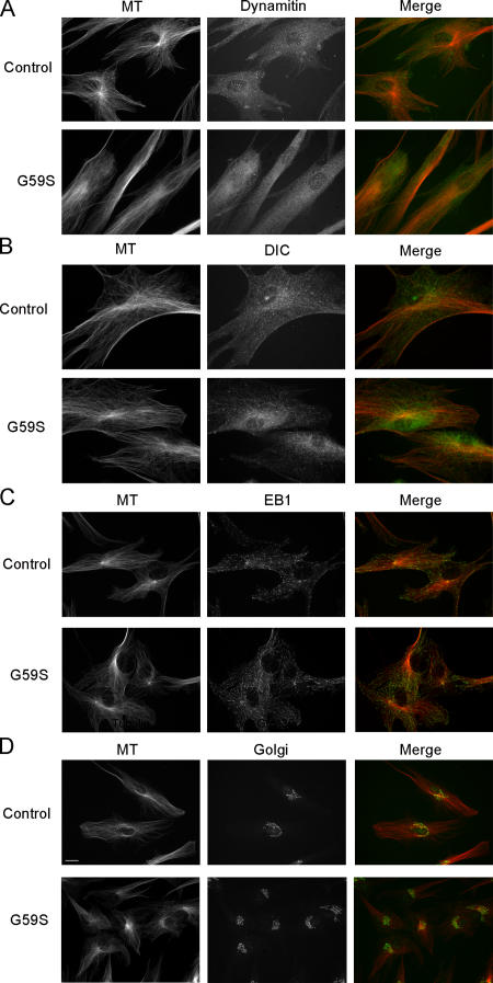
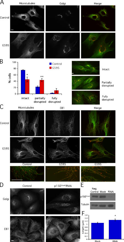
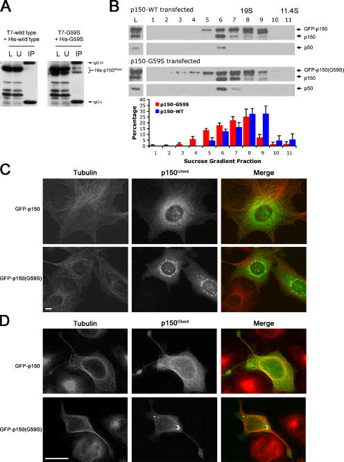
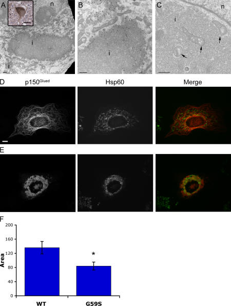
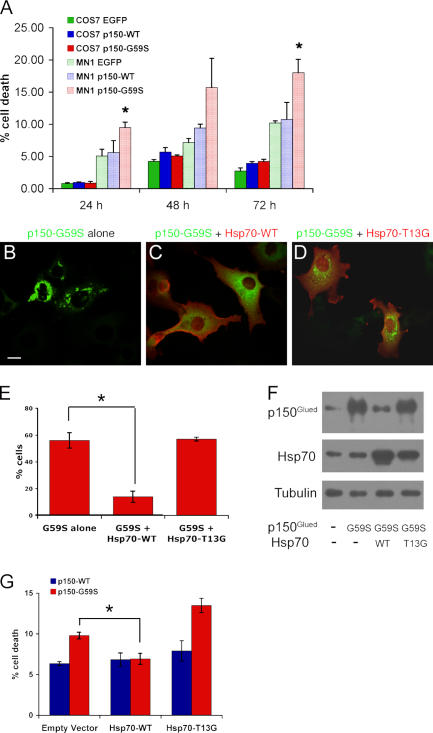
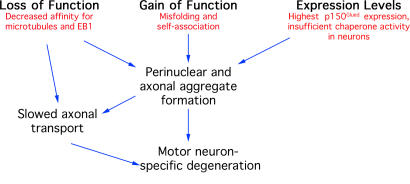
The microtubule motor cytoplasmic dynein and its activator dynactin drive vesicular transport and mitotic spindle organization. Dynactin is ubiquitously expressed in eukaryotes, but a G59S mutation in the p150Glued subunit of dynactin results in the specific degeneration of motor neurons. This mutation in the conserved cytoskeleton-associated protein, glycine-rich (CAP-Gly) domain lowers the affinity of p150Glued for microtubules and EB1. Cell lines from patients are morphologically normal but show delayed recovery after nocodazole treatment, consistent with a subtle disruption of dynein/dynactin function. The G59S mutation disrupts the folding of the CAP-Gly domain, resulting in aggregation of the p150Glued protein both in vitro and in vivo, which is accompanied by an increase in cell death in a motor neuron cell line. Overexpression of the chaperone Hsp70 inhibits aggregate formation and prevents cell death. These data support a model in which a point mutation in p150Glued causes both loss of dynein/dynactin function and gain of toxic function, which together lead to motor neuron cell death.


Figures

Figure 1.
Figure 1.
The G59S mutation impairs the binding of p150Glued to microtubules. (A) Schematic representation of p150Glued. Glycine 59 lies in the CAP-Gly microtubule binding domain. The 1–107 fragment contains the CAP-Gly domain, and the 1–333 fragment contains the CAP-Gly domain, an adjacent serine-rich domain (residues 111–191), and a small part of the first predicted coiled-coil domain. (B) Wild-type (WT) and G59S p150Glued were expressed in vitro and incubated with increasing concentrations of microtubules. The microtubule bound and unbound proteins were separated by centrifugation and visualized by SDS-PAGE and fluorography. The fraction bound, as determined by densitometry, was plotted against the concentration of tubulin ± SEM and fitted to a rectangular hyperbola. (C) COS7 cells were transfected with GFP-tagged wild-type (top) or G59S (bottom) p150Glued. Cells were fixed after 48 h and stained for GFP (green) and microtubules (red). Bar, 10 μm.
Figure 2.
Figure 2.
The G59S mutation impairs the binding of p150Glued to EB1 and to microtubule plus ends. (A) Affinity chromatography of in vitro–translated wild-type (WT) or G59S p150Glued (residues 1–333) over an EB1 column. Load (L), flow-through (F), wash (W), and eluate fractions (E1 and E2) were analyzed by SDS-PAGE and Western blot using a polyclonal antibody to p150Glued. There is less G59S p150Glued in the eluate fractions, in comparison to wild-type p150Glued, indicating a decrease in G59S p150Glued affinity for EB1. (B and C) Live cell fluorescence microscopy was used to observe the dynamics and localization of p150Glued in COS7 cells expressing low levels of GFP-tagged wild-type (B) and G59S (C) p150Glued. Bar, 10 μm. B and C show still images from Videos 1 and 2, respectively (available at http://www.jcb.org/cgi/content/full/jcb.200511068/DC1).
Figure 3.
Figure 3.
Expression of G59S p150Glued does not alter the integrity of the dynactin complex. (A) Quantification of levels of p150Glued RNA in lymphoblast and fibroblast cell lines derived from patients carrying the G59S mutation and unaffected controls, as measured by RT-PCR. n = 3. (B) Western blot analysis of dynactin expression levels in fibroblast and lymphoblast cell lines from control individuals (C) and patients heterozygous for the G59S mutation (P). Cell extracts were resolved by SDS-PAGE and probed for the dynactin subunit dynamitin, as well as DIC and actin. (C, right) An anti-p150Glued monoclonal antibody (mAb) directed against the CAP-Gly domain does not recognize in vitro–translated (IVT) G59S p150Glued. (left) An anti-p150Glued polyclonal antibody (pAb) recognizes both the wild-type and mutant in vitro–translated protein. (D) Relative levels of total and wild-type p150Glued expressed in fibroblasts isolated from patients carrying the G59S mutation. (E) Protein extracts from G59S and control fibroblast cell lines were sedimented on 5–25% sucrose gradients. The fractions were resolved by SDS-PAGE, and Western blot was performed using antibodies for the dynactin subunits p150Glued and DIC. There is no peak of dynactin subunits in the lower density fractions, indicating that these subunits are incorporated into the dynactin complex.
Figure 4.
Figure 4.
Dynein, dynactin, and EB1 are localized normally in cells heterozygous for the G59S mutation in p150Glued. Control fibroblasts and fibroblasts from patients heterozygous for the G59S mutation were stained with antibodies to tubulin (MT; red) and dynamitin (A), DIC (B), EB1 (C), and the Golgi marker GM130 (D). We observed no difference in morphology or microtubule organization in control and patient cells. There was a punctate, cytoplasmic localization of dynein and dynactin and microtubule tip localization of EB1 in both control and patient cells. The Golgi is intact and perinuclear in control and patient cells. Bar, 10 μm.
Figure 5.
Figure 5.
Cells heterozygous for the G59S mutation in p150Glued have delayed recovery after microtubule depolymerization. Nocodazole washout experiments were performed on patient and control fibroblasts. Cells were treated with nocodazole for 1 h, washed twice with PBS, and returned to normal growth media. (A) After 1 h of recovery, cells were fixed and stained for the cis-Golgi marker GM130 (red) and microtubules (green). Control cells have compact and perinuclear Golgi, but patient cells have partially disrupted Golgi at the same time point after drug washout. (B) Quantification of Golgi morphology after 1 h of recovery, ± SD (*, P < 0.05; **, P < 0.01). n = 3. (C) After 30 min of recovery, cells were fixed and stained for EB1 (red) and microtubules (green). Enlargements of merged images are shown at the bottom. Control cells show distinct tip localization of EB1, but patient cells show subtle mislocalization of EB1 along microtubules. (D) HeLa-M cells, either mock-transfected or transfected with small interfering RNA against p150Glued, stained with antibodies for EB1 or trans-Golgi marker 46. (E) Knockdown of p150Glued, compared with cells transfected with a fluorescein-labeled, nontargeting oligo or mock-transfected cells. (F) Quantification of the length of EB1 tails, ± SD (*, P < 0.05). Bars, 10 μm.
Figure 6.
Figure 6.
G59S p150Glued aggregates in vitro and in vivo. (A) His- and T7-tagged constructs of wild-type and G59S p150Glued were coexpressed in vitro. Immunoprecipitations were performed with anti-T7 antibody. The load (L), unbound (U), and immunoprecipitated (IP) fractions were resolved by SDS-PAGE and probed with antibodies to the His tag. The G59S construct, though not the wild type, runs as a doublet. We observed coimmunoprecipitation of the differentially tagged G59S constructs, indicating that the mutant protein self-associates. (B) Lysates from COS7 cells transfected with either GFP-tagged wild-type (WT) or G59S p150Glued constructs were sedimented on 5–25% sucrose gradients. The fractions were resolved by SDS-PAGE and probed for the dynactin subunits p150Glued and p50. The histogram indicates the mean percentage of protein in each fraction, as determined in three experiments, ± SEM. G59S p150Glued appears in the high-density fractions at a higher frequency than wild-type p150Glued, which indicates it is incorporated into a high–molecular weight complex. n = 4. (C) COS7 cells transfected with GFP-tagged wild-type or G59S p150Glued and fixed after 96 h. Bar, 10 μm. (D) MN1 cells transfected with wild-type or G59S p150Glued. Bar, 25 μm.
Figure 7.
Figure 7.
The p150Glued inclusions are associated with mitochondria. (A and B) Low-magnification (A) and high-magnification (B) electron micrographs of MN1 cells that have been transfected with GFP-labeled G59S p150Glued and immunolabeled with an antibody to GFP. Inclusions (i) and nuclei (n) are labeled. Bars, 500 nm. Inset, immunohis-tochemistry for DIC was performed on sections from the medulla of an affected patient. Bar, 10 μm. (C) High-magnification electron micrograph of a COS7 cell that has been transfected with GFP-labeled G59S p150Glued and fixed with glutaraldahyde. No membrane surrounds the inclusion; the visible membrane is a nuclear envelope. Arrows indicate mitochondria surrounding and within G59S p150Glued protein inclusions. Bar, 500 nm. (D and E) COS7 cells transfected with GFP-tagged wild-type (D) or G59S (E) p150Glued and fixed and stained using antibodies for p150Glued and Hsp60. Bar, 10 μm. (F) Quantification of area of cells containing mitochondria in arbitrary units, ± SEM (*, P < 0.05). Wild type, n = 8; G595, n = 12.
Figure 8.
Figure 8.
Overexpression of Hsp70 decreases both aggregation of G59S p150Glued and MN1 cell death. (A) Quantitation of cell death after transfection with wild-type (WT) or G59S p150Glued or EGFP alone, as determined by PI exclusion. Values represent mean percentage of cell death in three sets of transfections, ± SEM (*, P < 0.05). n = 3. (B–D) Representative images of cells transfected with G59S p150Glued alone (B), G59S p150Glued and wild-type Hsp70 (C), or G59S p150Glued and T13G Hsp70 (D). Cells were stained with antibodies for p150Glued (green) and the hemagglutinin tag on the Hsp70 constructs (red). Bar, 10 μm. (E) Quantitation of the percentage of COS7 cells containing aggregates, ± SD (*, P < 0.05). n = 2. (F) Expression levels of p150Glued and Hsp70 in lysates from cells that have been mock-transfected, transfected with G59S alone, and transfected with wild-type or T13G Hsp70. Tubulin was used as a loading control. (G) Quantitation of mean percentage of cell death in MN1 cells 48 h after transfection with wild-type and G59S p150Glued and wild-type and T13G Hsp70 or empty vector, ± SEM (*, P < 0.05). Cotransfection of G59S p150Glued and wild-type Hsp70 protects MN1 cells from death, but cotransfection of G59S p150Glued with T13G Hsp70 or empty vector does not. n = 3.
Figure 9.
Figure 9.
Proposed mechanism of G59S p150Glued–mediated motor neuron toxicity. G59S p150Glued expression leads to motor neuron toxicity through three intersecting pathways that lead to cell death. The mutation causes impaired microtubule and EB1 binding, which leads to disrupted dynein/dynactin-based transport. In addition, the mutation causes misfolding of the CAP-Gly domain, which leads to aberrant self-association. The large proportion of unbound p150Glued, along with the high expression levels of p150Glued in neurons, leads to a high cytosolic concentration of misfolded mutant protein resulting in aggregates specifically in neurons. This gain of function may induce further impairment in axonal transport, either by physical blockage of the axon or by sequestration of dynein and dynactin, leading to motor neuron–specific degeneration.

References

    1. Barral, J.M., S.A. Broadley, G. Schaffar, and F.U. Hartl. 2004. Roles of molecular chaperones in protein misfolding diseases. Semin. Cell Dev. Biol. 15:17–29. - PubMed
    1. Brooks, B.P., D.E. Merry, H.L. Paulson, A.P. Lieberman, D.L. Kolson, and K.H. Fischbeck. 1998. A cell culture model for androgen effects in motor neurons. J. Neurochem. 70:1054–1060. - PubMed
    1. Bruijn, L.I., T.M. Miller, and D.W. Cleveland. 2004. Unraveling the mechanisms involved in motor neuron degeneration in ALS. Annu. Rev. Neurosci. 27:723–749. - PubMed
    1. Burkhardt, J.K., C.J. Echeverri, T. Nilsson, and R.B. Vallee. 1997. Overexpression of the dynamitin (p50) subunit of the dynactin complex disrupts dynein-dependent maintenance of membrane organelle distribution. J. Cell Biol. 139:469–484. - PMC - PubMed
    1. Carson, J.H., H. Cui, and E. Barbarese. 2001. The balance of power in RNA trafficking. Curr. Opin. Neurobiol. 11:558–563. - PubMed

Publication types

MeSH terms