. 2006 Mar 7;103(10):3569-74.
doi: 10.1073/pnas.0600332103. Epub 2006 Feb 27.

A DEAD-box protein alone promotes group II intron splicing and reverse splicing by acting as an RNA chaperone

Affiliations

A DEAD-box protein alone promotes group II intron splicing and reverse splicing by acting as an RNA chaperone

Sabine Mohr et al. Proc Natl Acad Sci U S A. .

Abstract

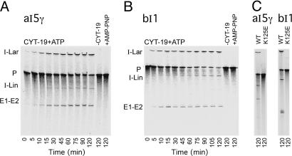
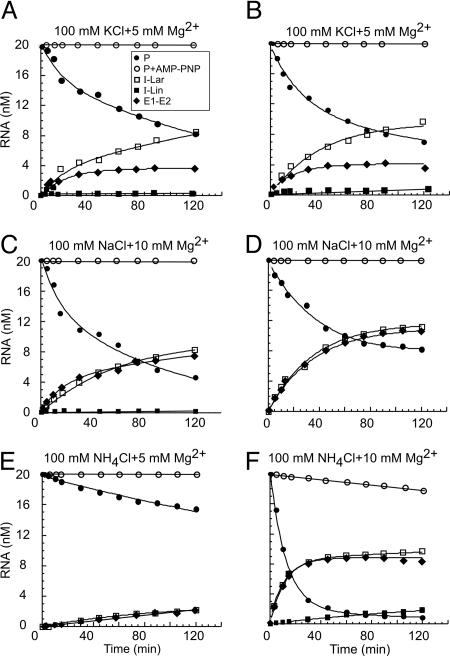
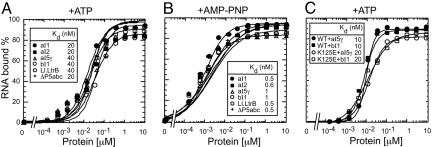
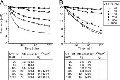
Group II intron RNAs self-splice in vitro but only at high salt and/or Mg2+ concentrations and have been thought to require proteins to stabilize their active structure for efficient splicing in vivo. Here, we show that a DEAD-box protein, CYT-19, can by itself promote the splicing and reverse splicing of the yeast aI5gamma and bI1 group II introns under near-physiological conditions by acting as an ATP-dependent RNA chaperone, whose continued presence is not required after RNA folding. Our results suggest that the folding of some group II introns may be limited by kinetic traps and that their active structures, once formed, do not require proteins or high Mg2+ concentrations for structural stabilization. Thus, during evolution, group II introns could have spliced and transposed by reverse splicing by using ubiquitous RNA chaperones before acquiring more specific protein partners to promote their splicing and mobility. More generally, our results provide additional evidence for the widespread role of RNA chaperones in folding cellular RNAs.


Conflict of interest statement

Conflict of interest statement: No conflicts declared.

Figures

Fig. 1.
Fig. 1.
CYT-19-dependent splicing of group II introns aI5γ and bI1. (A and B) Splicing time courses were done by incubating 32P-labeled RNA substrate (20 nM) containing the intron and flanking exon sequences with purified CYT-19 protein (500 nM) plus 1 mM ATP in reaction medium containing 100 mM KCl/5 mM MgCl2 at 30°C. Products were analyzed in a denaturing 4% polyacrylamide gel. Control lanes (Right) show RNA substrate incubated without CYT-19 (−CYT-19) or with CYT-19 plus 1 mM AMP-PNP (+AMP-PNP) for 120 min. (C) Splicing reactions with wild-type CYT-19 (WT) and mutant K125E were done as above for 120 min. E1-E2, ligated exons; I-Lar, intron lariat; I-Lin, linear intron; P, precursor RNA. The I-Lin band may contain a mixture of linear intron and broken lariat RNA. The closely spaced doublet for bI1 ligated exons seen previously (21) likely is caused by a premature transcription stop.
Fig. 2.
Fig. 2.
Representative kinetic analysis of CYT-19-dependent splicing of aI5γ and bI1. Splicing time courses for aI5γ (A, C, and E) and bI1 (B, D, and F) were done with 500 nM CYT-19, 20 nM 32P-labeled precursor RNA, and 1 mM ATP or AMP-PNP at 30°C in reaction media containing 100 mM KCl (A and B), NaCl (C and D), or NH4Cl (E and F), and 5 or 10 mM MgCl2, as indicated. The plots show the disappearance of precursor RNA and appearance of products as a function of time. In some reactions (e.g., aI5γ in NaCl and bI1 in NH4Cl), precursor RNA degradation at long-time points leads to a 20–30% discrepancy in molar ratios of substrate and products. Differential susceptibility to trace nuclease activity may also account for the finding that in the KCl reactions lariat RNA continues to accumulate at long time points, whereas ligated exons plateau. I-Lin was not detectable in reactions D and E. No CYT-19-promoted splicing was observed for either aI5γ or bI1 in reaction media containing 50 or 100 mM (NH4)2SO4 (an optimal salt for aI5γ self-splicing; ref. 20) and 5 or 10 mM MgCl2 at 30°C (data not shown). Abbreviations are as in Fig. 1.
Fig. 3.
Fig. 3.
Equilibrium-binding assays. The indicated 32P-labeled RNAs (5 pM) were incubated with increasing concentrations of CYT-19 protein for 90 min at 30°C, and then filtered through nitrocellulose to bind RNA–protein complexes. A and B compare the binding of a group I intron (T. thermophila LSU-ΔP5abc intron) and the indicated group II introns RNAs in reaction medium containing 100 mM KCl/5 mM MgCl2 with 1 mM ATP or 1 mM AMP-PNP, respectively. C compares the binding of wild-type CYT-19 (WT) and mutant K125E to aI5γ and bI1 in reaction medium containing 100 mM KCl/5 mM MgCl2 plus 1 mM ATP. The plots show the percent of the input RNA bound to a nitrocellulose filter as a function of CYT-19 concentration. Essentially identical binding curves were obtained when RNA and protein were incubated for 10 or 30 min (data not shown).
Fig. 4.
Fig. 4.
CYT-19-concentration dependence of group II intron splicing. Splicing time courses with aI5γ (A) and bI1 (B) were done with 20 nM precursor RNA and 20 nM to 1 μM CYT-19 plus 1 mM ATP in reaction media containing 100 mM KCl/5 mM Mg2+ at 30°C. The data were best-fit to equations with one or two exponentials, and calculated rate constants for the single phase (k1) or the fast and slow phases (k1 and k2, respectively) are summarized beneath the plots. The numbers in parentheses indicate the proportion of total RNA spliced in each phase. The I-Lin band, which could contain either linear intron or broken lariat RNA, was not detectable at or below 200 nM CYT-19 for either intron, but increased to ≈4% of the total product at 500 mM CYT-19 and is equimolar with I-lar at 1 μM CYT-19. This increase most likely reflects increased breakage of lariat RNA caused by contaminating nucleases at high protein concentrations.
Fig. 5.
Fig. 5.
Reverse-branching and reverse-splicing reactions. (A and B) Reactions with bI1. Gel-purified intron lariat RNA was preincubated under the conditions indicated below, before initiating the reactions by adding 5′-labeled (star) RNA oligonucleotide corresponding to the 5′ exon (E1) (reverse branching) or ligated exons (E1–E2) (reverse splicing). Lanes 1–3, lariat RNA preincubated for 90 min without (lane 1) or with CYT-19 plus ATP (lane 2) or CYT-19 plus AMP-PNP (lane 3). Lanes 4 and 5, lariat RNA preincubated with CYT-19 plus ATP for 1 h, then split with halves incubated another 30 min without (lane 4) or with protease K (Prot-K) plus SDS (lane 5). Lane 6, lariat RNA incubated for 1 h with CYT-19 plus AMP-PNP and then for 30 min with protease K plus SDS. (C) Reverse branching of the L. lactis Ll.LtrB intron. Lanes 1 and 2, lariat RNA preincubated for 50 min without (lane 1) or with LtrA maturase (lane 2). Lanes 3 and 4, lariat RNA preincubated with LtrA maturase for 20 min, then split with halves incubated for 30 min without (lane 3) or with protease K (Prot-K) plus SDS (lane 4). Lane 5, lariat RNA preincubated for 20 min and then incubated for 30 min with protease K plus SDS. All incubations were at 30°C. Products were analyzed in denaturing 4% (upper gel) and 10% (lower gel) polyacrylamide gels. Schematics of the reverse-branching and reverse-splicing reactions are shown at the bottom.

References

    1. Michel M., Ferat J. L. Biochemistry. 1995;64:435–461. - PubMed
    1. Lehmann K., Schmidt U. Crit. Rev. Biochem. Mol. Biol. 2003;38:249–303. - PubMed
    1. Pyle A. M., Lambowitz A. M. In: The RNA World. 3rd Ed. Gesteland R. F., Atkins J. F., Cech T. R., editors. Plainview, NY: Cold Spring Harbor Lab Press; 2006. pp. 469–505.
    1. Lambowitz A. M., Caprara M. G., Zimmerly S., Perlman P. S. In: The RNA World. 2nd Ed. Gesteland R. F., Atkins J. F., Cech T. R., editors. Plainview, NY: Cold Spring Harbor Lab Press; 1999. pp. 451–485.
    1. Matsuura M., Saldanha R., Ma H., Wank H., Yang J., Mohr G., Cavanagh S., Dunny G. M., Belfort M., Lambowitz A. M. Genes Dev. 1997;11:2910–2924. - PMC - PubMed

Publication types

MeSH terms

LinkOut - more resources