. 2006 Mar 7;103(10):3610-4.
doi: 10.1073/pnas.0600239103. Epub 2006 Feb 27.

Electron trap for DNA-bound repair enzymes: a strategy for DNA-mediated signaling

Affiliations

Electron trap for DNA-bound repair enzymes: a strategy for DNA-mediated signaling

Eylon Yavin et al. Proc Natl Acad Sci U S A. .

Abstract

Despite a low copy number within the cell, base excision repair (BER) enzymes readily detect DNA base lesions and mismatches. These enzymes also contain [Fe4S4] clusters, yet a redox role for these iron cofactors had been unclear. Here, we provide evidence that BER proteins may use DNA-mediated redox chemistry as part of a signaling mechanism to detect base lesions. By using chemically modified bases, we show electron trapping on DNA in solution with bound BER enzymes by electron paramagnetic resonance (EPR) spectroscopy. We demonstrate electron transfer from two BER proteins, Endonuclease III (EndoIII) and MutY, to modified bases in DNA containing oxidized nitroxyl radical EPR probes. Electron trapping requires that the modified base is coupled to the DNA pi-stack, and trapping efficiency is increased when a noncleavable MutY substrate analogue is located distally to the trap. These results are consistent with DNA binding leading to the activation of the repair proteins toward oxidation. Significantly, these results support a mechanism for DNA repair that involves DNA-mediated charge transport.


Conflict of interest statement

Conflict of interest statement: No conflicts declared.

Figures

Fig. 1.
Fig. 1.
Proposed model for long-range DNA signaling between BER enzymes using DNA-mediated CT to detect base lesions. In the absence of DNA, the [Fe4S4]2+ clusters in the BER enzymes are robust to oxidation. Binding to DNA activates the BER enzymes to oxidation, facilitating DNA-mediated CT to an alternate protein bound at a distal site. Reduction of the distally bound protein then promotes its dissociation. Binding of a protein near a base lesion, however, precludes DNA-mediated CT; hence, the protein remains localized in the vicinity of the lesion and can diffuse processively to the site to excise the base. DNA CT therefore promotes a redistribution of the repair enzymes near sites in need of repair.
Fig. 2.
Fig. 2.
Electron trapping on the DNA duplex. (a) The methodology used. The stable nitroxyl radical is covalently attached to a uridine base on the DNA duplex. This organic radical may be readily monitored by EPR at ambient temperature. Mild oxidation with Ir(IV) of the nitroxyl radical generates the diamagnetic N-oxo-ammonium species. Upon protein binding, electron trapping on the DNA then can be monitored by the reappearance of the EPR signal of the nitroxyl radical. (b) The DNA assemblies and modified bases used.
Fig. 3.
Fig. 3.
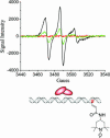
EPR spectra before and after addition of MutY (Left) and EndoIII (Right) are shown for DNA duplexes (7 μM) initially treated with IrCl6−2 (60 μM). Spectra shown are before oxidation (black), after oxidation before protein addition (red), and then after protein addition (green). Protein concentrations are 12 μM for MutY and 14 μM for EndoIII.
Fig. 4.
Fig. 4.
EPR spectra of a DNA duplex (6.6 μM) containing the nitroxyl radical-modified base separated by 19 base pairs from a GZ base pair after (red) treatment with IrCl6−2 (60 μM) followed by the addition of 6.2 μM MutY (blue). An increase in the signal intensity is observed in comparison with an identical duplex lacking the GZ base pair (green).
Fig. 5.
Fig. 5.
EPR spectra of a DNA duplex (7 μM) containing a poorly conjugated spin label (spin label 2, black) initially treated with 60 μM IrCl6−2 (red) followed by the addition of 14 μM EndoIII (green).

References

    1. O’Neill M. A., Barton J. K. In: Charge Transfer in DNA: From Mechanism to Application. Wagenknecht H.-A., editor. Weinheim, Germany: Wiley; 2005. pp. 27–75.
    1. Hall D. B., Holmlin R. E., Barton J. K. Nature. 1996;382:731–735. - PubMed
    1. Nakatani K., Dohno C., Saito I. J. Am. Chem. Soc. 1999;121:10854–10855.
    1. Meggers E., Kusch D., Spichty M., Wille U., Giese B. Angew. Chem. Int. Ed. 1998;37:460–462. - PubMed
    1. Gasper S. M., Schuster G. B. J. Am. Chem. Soc. 1997;119:12762–12771.

Publication types