. 2006 Mar 7;103(10):3752-7.
doi: 10.1073/pnas.0506977103. Epub 2006 Feb 27.

Reprogramming metastatic melanoma cells to assume a neural crest cell-like phenotype in an embryonic microenvironment

Affiliations

Reprogramming metastatic melanoma cells to assume a neural crest cell-like phenotype in an embryonic microenvironment

Paul M Kulesa et al. Proc Natl Acad Sci U S A. .

Abstract

Human metastatic melanoma cells express a dedifferentiated, plastic phenotype, which may serve as a selective advantage, because melanoma cells invade various microenvironments. Over the last three decades, there has been an increased focus on the role of the tumor microenvironment in cancer progression, with the goal of reversing the metastatic phenotype. Here, using an embryonic chick model, we explore the possibility of reverting the metastatic melanoma phenotype to its cell type of origin, the neural-crest-derived melanocyte. GFP-labeled adult human metastatic melanoma cells were transplanted in ovo adjacent to host chick premigratory neural crest cells and analyzed 48 and 96 h after egg reincubation. Interestingly, the transplanted melanoma cells do not form tumors. Instead, we find that transplanted melanoma cells invade surrounding chick tissues in a programmed manner, distributing along host neural-crest-cell migratory pathways. The invading melanoma cells display neural-crest-cell-like morphologies and populate host peripheral structures, including the branchial arches, dorsal root and sympathetic ganglia. Analysis of a melanocyte-specific phenotype marker (MART-1) and a neuronal marker (Tuj1) revealed a subpopulation of melanoma cells that invade the chick periphery and express MART-1 and Tuj1. Our results demonstrate the ability of adult human metastatic melanoma cells to respond to chick embryonic environmental cues, a subset of which may undergo a reprogramming of their metastatic phenotype. This model has the potential to provide insights into the regulation of tumor cell plasticity by an embryonic milieu, which may hold significant therapeutic promise.


Conflict of interest statement

Conflict of interest statement: No conflicts declared.

Figures

Fig. 1.
Fig. 1.
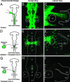
Transplanted metastatic melanoma cells invade chick cranial NCC migratory pathways and destinations. Chick embryos (6- to 8-somite stage) were injected with a lipophilic dye, DiI, into the rostral neural tube to label premigratory NCCs. Adult human metastatic melanoma cells (C8161) were then transplanted into specific cranial neural tube locations in the host chick embryos, one small block of melanoma cells per host chick embryo. Eggs were resealed and reincubated for 48 h. (AC) GFP-labeled melanoma cells, transplanted as a small clump of cells into the chick midbrain (m) region invade host tissue and spread out toward the ba1 and eye (e). (B and C) The GFP-labeled melanoma cells (green) migrate with the host chick NCCs (red; asterisk) along the stereotypical NCC pathway. (DF) GFP-labeled melanoma cells (green) transplanted into r2 spread out to populate ba1 (arrow), together with host chick NCCs (red). (GI) GFP-labeled melanoma cells (green; arrowhead) transplanted into r6 reach the ba4 with host chick NCCs (red). A total of 128 transplantations were performed, one transplant per embryo. The otic vesicle (ov) is labeled. [Scale bars, 100 μm (B and C) and 50 μm (E, F, H, and I).]
Fig. 2.
Fig. 2.
Transplanted metastatic melanoma cells and chick NCCs share in vivo cell morphologies along host NCC migratory pathways. The images demonstrate three commonly observed cell morphologies of host chick NCCs and human melanoma cells transplanted into chick embryos. (A and B) The first example shows typical host chick NCCs (A) and transplanted melanoma cells (B) displaying long filopodial extensions. Both cell types display a bipolar phenotype. (C and D) The second example shows two individual NCCs (C) and melanoma cells (D) connected by a thin (1–2 μm) filopodial connection. (E and F) The third example shows host chick NCCs (E) and melanoma cells (F) displaying collective cell migratory structures in linear chain-like arrays, with the melanoma cell chains typically longer than the NCCs chains. The NCCs (A, C, and E) are labeled with a fluorescent protein construct Gap43-EGFP and double-labeled in E with H2B-mRFP. [Scale bars, 10 μm (AE) and 50 μm (F).] (G and H) The graphs represent the number of hairy/round vs. bipolar-shaped cells counted after analyzing migrating neural crest (G) and metastatic melanoma (H) cells invading the chick embryo. (G) In a typical fluorescently labeled chick embryo (n = 9 embryos; n = 361 NCCs counted), the blue bars represent NCCs in the middle of NCC migratory streams vs. in the branchial arches (red).
Fig. 3.
Fig. 3.
Metastatic melanoma cells transplanted into the chick trunk neural tube invade DRG and SG sites. (A) Metastatic melanoma cells are transplanted into the trunk neural tube (arrow) in a typical chick embryo host with 9–11 somites and (B) incubated for ≈48 h. The location of the melanoma cell transplant is shown by the green square and arrow in a typical embryo. (C) A transverse view through the trunk region of a typical chick embryo in the region of the transplanted melanoma cells (green) 48 h after transplantation and incubation (the outline surrounds the neural tube). A thick transverse section was cut through the embryo (200 μm) by using a razor blade and laid flat to reveal ventral tissues. The melanoma cells (green) have migrated from the dorsal neural tube to the DRG and further ventral. Melanoma cells also appear along a dorsolateral route. (D) A sagittal view through a typical chick embryo in the region of the transplanted melanoma cells (green) 48 h after transplantation. The embryo was cut in half down the anterior–posterior axis and laid flat to reveal ventral tissues. The background staining is HNK-1 (red), showing the host chick trunk NCCs migrating in stripes through the rostral, but not the caudal, portions of somites. The melanoma cells (green) appear to colocalize with the HNK-1-positive (red) stripes and migrate to the DRG and SG (E) The melanoma cells (green) target the forming SG and colocalize with the HNK-1-labeled trunk NCCs (red). A total of 40 transplantations were performed, one transplant per embryo. (Scale bars, 50 μm.)
Fig. 4.
Fig. 4.
Transplantation of metastatic melanoma cells into odd-numbered chick rhombomeres after ablation of host chick premigratory NCCs. In normal chick embryos, DiI-labeled cranial NCCs (AC) are sculpted into three stereotypical migratory streams that extend from the rhombomeres (r), r1 and r2, r4, and r6. (B and C) After 36 h, the streams lead to the branchial arch destinations: NCCs from r3, r4, and r5 form into the r4 migratory stream (C) that targets the ba2. (DF) Host mid-r3 to mid-r5 premigratory NCCs are ablated in 9-somite chick embryos after a time when host chick NCCs regenerate. (E) Melanoma cells transplanted into r4 form a dense migratory stream along the stereotypical NCC migratory pathway and (F) populate the ba2 and ba3 and the region near ba1 when viewed after 48 h. (GI) At 48 h after melanoma cells are transplanted into r3, they do not form into a dense migratory stream to invade the host chick tissue (H) but remain fairly confined to the neural tube. (I) Only a small number of melanoma cells emerge from the neural tube. A total of 72 ablations plus transplantations were performed, one transplant per embryo. [Scale bars, 100 μm (B, C, E, and F); and the scale bars in E and F can be used for H and I, respectively.]
Fig. 5.
Fig. 5.
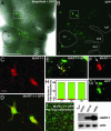
MART-1 expression of transplanted melanoma cells after invading host chick embryonic tissues. (A) A typical chick embryo 48 h after transplantation of GFP-labeled (green) metastatic melanoma cells and reincubation. (A and B) The GFP-labeled melanoma cells (green) invade the chick host periphery around the otic vesicle (ov, circled) and target the ba1–ba3 (outlined). (AD) After fixation and staining for MART-1, the box surrounds two individual melanoma cells that are shown magnified in C and D positive for MART-1 expression. (EH) Further analysis of invading melanoma cells in tissue sections shows that a number of melanoma cells express MART-1. The location of a typical tissue section is shown by a dotted line in A. (H) The bar graph plots the percent number of MART-1-positive melanoma cells (red) vs. the total number of GFP-labeled metastatic melanoma cells (green) transplanted into r1, r4, and r6. (I) Before transplantation of melanoma cells (C8161) into the chick embryo, melanoma cells were stained for MART-1. (J) Western blot analysis for the expression of MART-1 in cell lysates of the human melanoma cell lines C8161 (highly aggressive and metastatic) and C81-61 (poorly aggressive) as well as the human melanocyte cell line HEMn. Fifteen micrograms of protein were loaded per lane of each sample, and Western analysis revealed the presence of MART-1 in C81-61 cells and HEMn melanocytes. An equal loading of the samples is demonstrated by probing the same blot for actin. [Scale bars, 200 μm (A and B), 15 μm (C and D).]

Similar articles

Cited by

References

    1. Mintz B., Illmensee K. Proc. Natl. Acad. Sci. USA. 1975;72:3585–3589. - PMC - PubMed
    1. Dolberg D. S., Bissell M. J. Nature. 1984;309:552–556. - PubMed
    1. Erickson C. A., Tosney K. W., Weston J. A. Dev. Biol. 1980;77:142–156. - PubMed
    1. Kenny P. A., Bissell M. J. Int. J. Cancer. 2003;107:688–695. - PMC - PubMed
    1. Hochedlinger K., Blelloch R., Brennan C., Yamada Y., Kim M., Chin L., Jaenisch R. Genes Dev. 2004;18:1875–1885. - PMC - PubMed

Publication types

MeSH terms

LinkOut - more resources