Comparative Study
. 2006 Apr 28;281(17):12069-80.
doi: 10.1074/jbc.M511621200. Epub 2006 Feb 27.

Endocytic function of von Hippel-Lindau tumor suppressor protein regulates surface localization of fibroblast growth factor receptor 1 and cell motility

Affiliations
Comparative Study

Endocytic function of von Hippel-Lindau tumor suppressor protein regulates surface localization of fibroblast growth factor receptor 1 and cell motility

Tien Hsu et al. J Biol Chem. .

Abstract

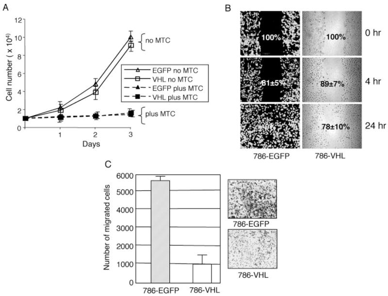
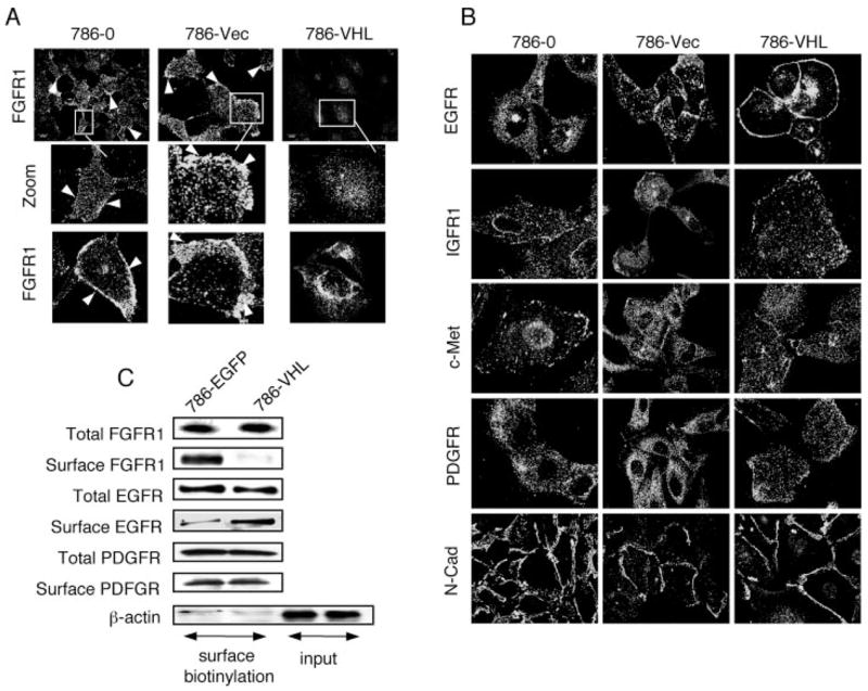
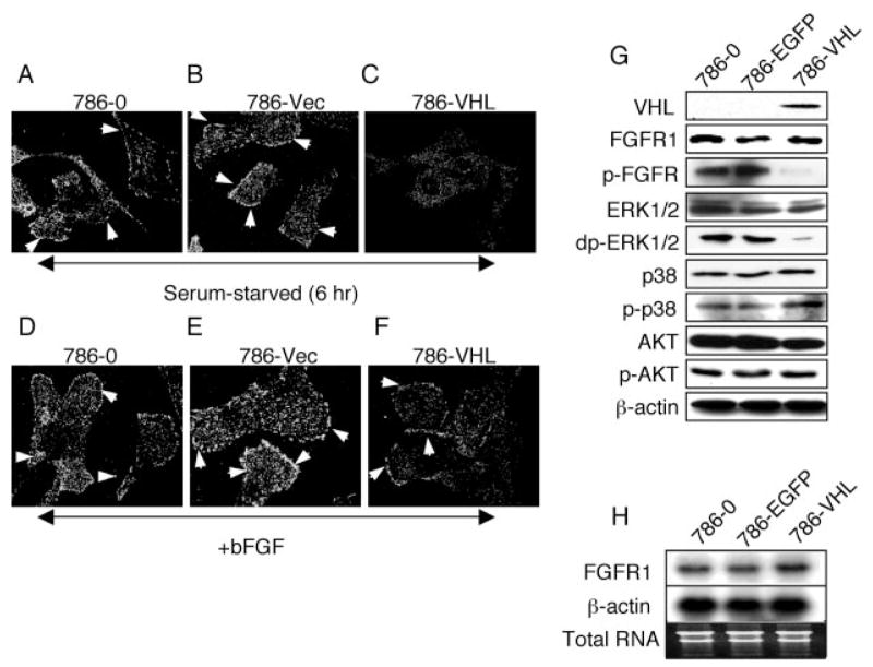
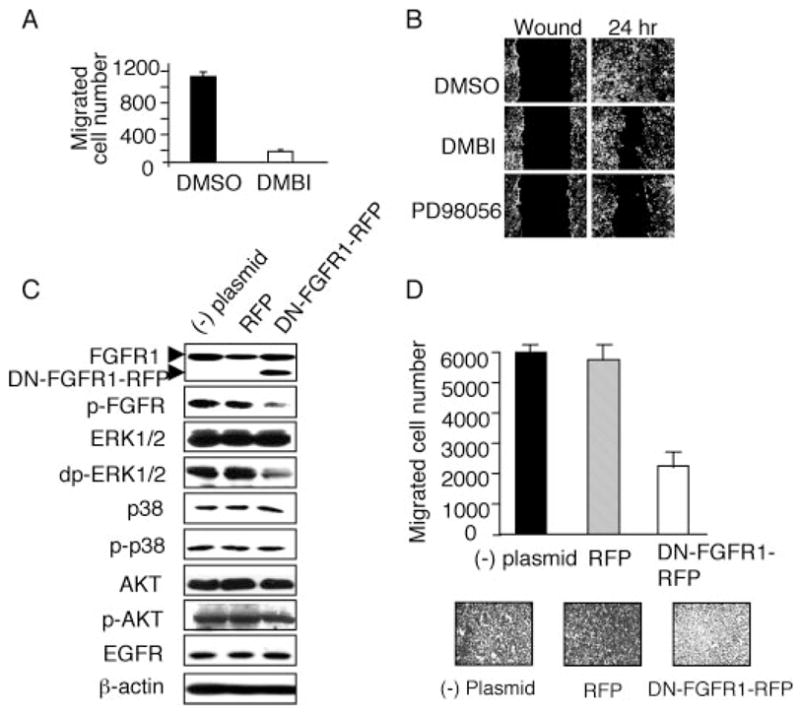
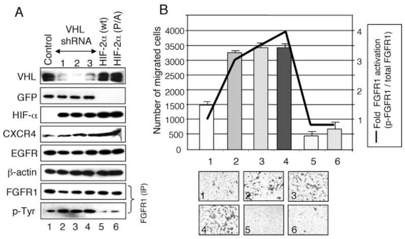
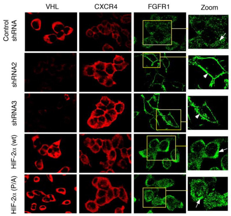
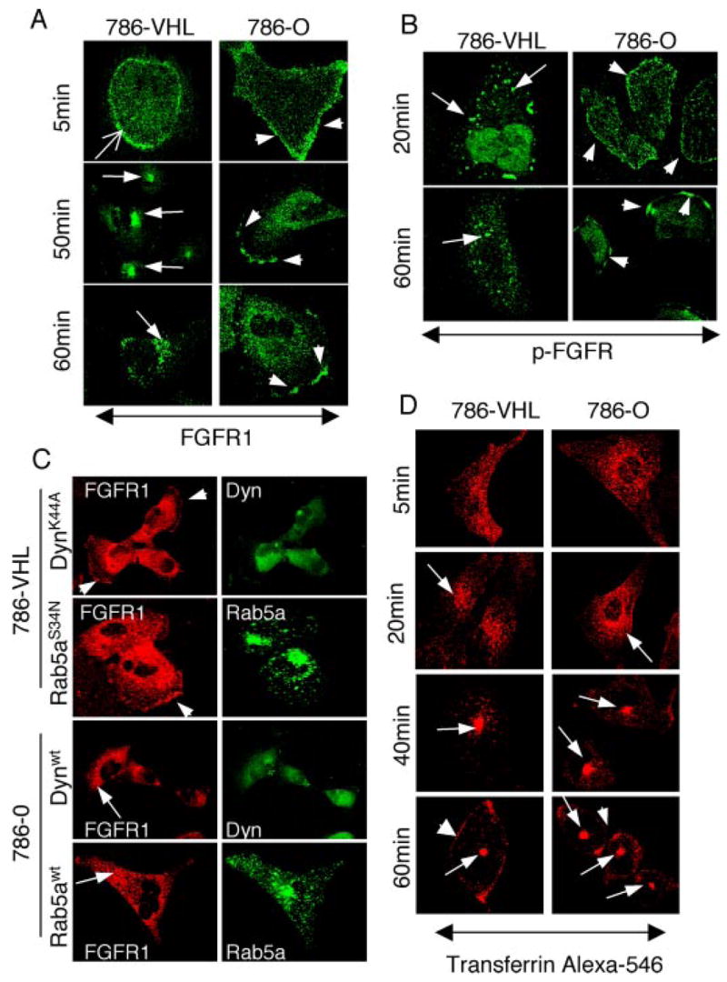
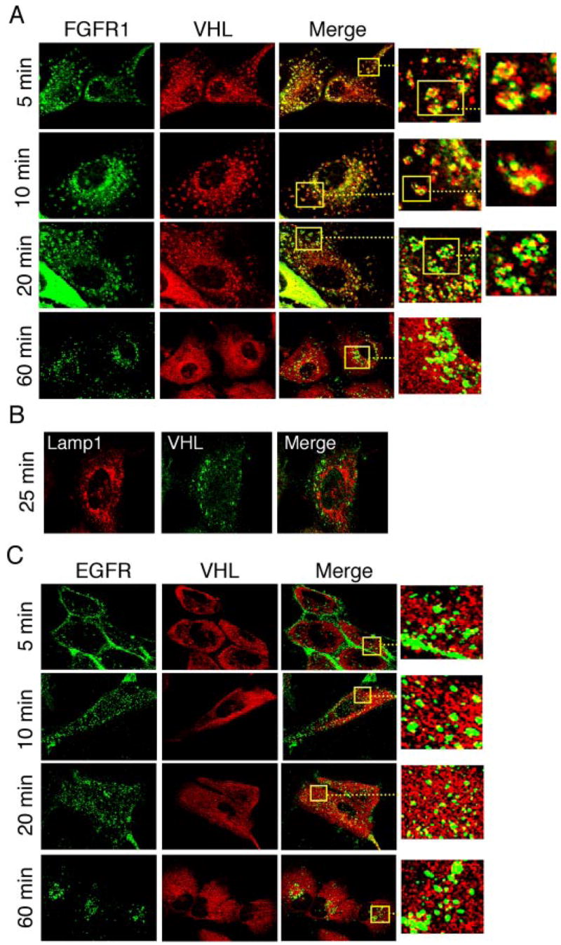
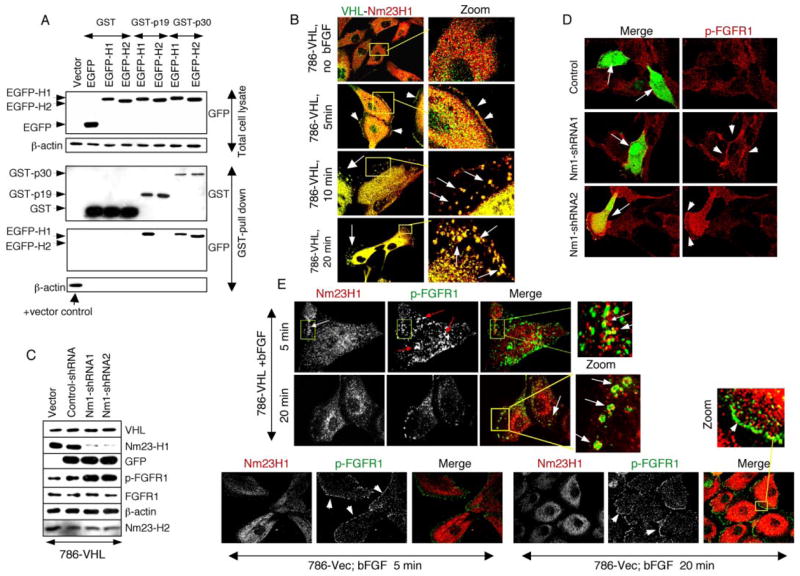
The tumor suppressor VHL (von Hippel-Lindau protein) serves as a negative regulator of hypoxia-inducible factor-alpha subunits. However, accumulated evidence indicates that VHL may play additional roles in other cellular functions. We report here a novel hypoxia-inducible factor-independent function of VHL in cell motility control via regulation of fibroblast growth factor receptor 1 (FGFR1) endocytosis. In VHL null tumor cells or VHL knock-down cells, FGFR1 internalization is defective, leading to surface accumulation and abnormal activation of FGFR1. The enhanced FGFR1 activity directly correlates with increased cell migration. VHL disease mutants, in two of the mutation hot spots favoring development of renal cell carcinoma, failed to rescue the above phenotype. Interestingly, surface accumulation of the chemotactic receptor appears to be selective in VHL mutant cells, since other surface proteins such as epidermal growth factor receptor, platelet-derived growth factor receptor, IGFR1, and c-Met are not affected. We demonstrate that 1) FGFR1 endocytosis is defective in the VHL mutant and is rescued by reexpression of wild-type VHL, 2) VHL is recruited to FGFR1-containing, but not EGFR-containing, endosomal vesicles, 3) VHL exhibits a functional relationship with Rab5a and dynamin 2 in FGFR1 internalization, and 4) the endocytic function of VHL is mediated through the metastasis suppressor Nm23, a protein known to regulate dynamin-dependent endocytosis.


Figures

FIGURE 1
FIGURE 1. Re-expression of VHL inhibits cell motility in VHL mutant cells
A, cell counts were obtained for 786-EGFP and 786-VHL stable cells grown in 1% serum conditions in the presence (plus MTC) or absence (no MTC) for the indicated duration. No significant difference in cell proliferation was observed between the two cell types in three independent experiments done in triplicate. Treatment with MTC inhibited cell proliferation in both cell types to a similar extent. B, 90% confluent cell monolayers in a 6-well culture dish were wounded with a 1000-μl pipette tip. Wound healing was followed for 786-EGFP and 786-VHL cells for 24 h. Representative images of four experiments are shown. 786-EGFP cells (left) are visualized with fluorescence, whereas 786-VHL cells (right) are with transmitted optics. The numbers within panels (with S.D. values) indicate the percentage gap left between the wound edges (indicating wound closure) at the specified time points. C, Boyden chamber assays for chemotactic cell migration showing an ~6-fold decrease of cell numbers migrated to the underside of the barrier membrane (p < 0.005) in 786-VHL compared with 786-EGFP cells in 12 h. The insets show representative images of migrated cells from six experiments done in triplicate. The error bars indicate S.D. (n = 18).
FIGURE 2
FIGURE 2. Surface accumulation of FGFR1 in VHL null cells
A, FGFR1 antibody staining of 786-O parental cells, 786-O transfected with empty pCDNA3.1 vector (786-Vec), and 786-O transfected with pCMV-VHL (786-VHL). Surface accumulation of FGFR1 (arrowheads) is observed in 786-O and 786-Vec but not in 786-VHL cells. Views of large populations (upper panels) and individual cells (insets and lower panels) are shown. B, 786-O, 786-Vec, and 786-VHL cells were stained with antibodies indicated on the left. There is no increase in surface accumulation of these membrane proteins in the VHL(−) cells. C, 786-EGFP and 786-VHL cells are labeled with membrane-impermeable sulfo-NHS-LC-biotin. After immunoprecipitation with specific rabbit polyclonal antibodies, the total levels of each receptor were detected using mouse monoclonal antibodies, and the cell surface fraction was distinguished using streptavidin-horseradish peroxidase. For β-actin, cell lysates were bound to streptavidin-agarose, and the eluted proteins were checked for the greatly reduced presence of β-actin (surface biotinylation), as compared with the input lanes (probed for β-actin as loading controls) that represent one-tenth of the lysates used for immunoprecipitation. Consistent with immunostaining results (A), there is significantly lower surface localization of FGFR1 in 786-VHL cells compared with 786-EGFP cells. EGFR surface localization is slightly lower in 786-EGFP cells. PDGFR shows no change.
FIGURE 3
FIGURE 3. Prolonged accumulation of activated FGFR1 in VHL mutant cells
786-O (A), 786-Vec (B), and 786-VHL cells (C), as described in the legend to Fig. 2A, were serum-starved for 6 h. Cells were fixed and stained for phosphorylated p-FGFR (indicating activation). 786-O (D), 786-Vec (E), and 786-VHL cells (F) were serum-starved for 12 h and then incubated with bFGF and heparin for 2 h at 4 °C. Cells were then shifted to 37 °C and processed for immunostaining for p-FGFR 5 min later. p-FGFR persists on the cell surface at 6 h after starvation in 786-O and 786-Vec cells (arrowheads in A and B), whereas in 786-VHL cells, very little p-FGFR1 is detected (C). The addition of bFGF plus heparin further increased surface p-FGFR level in 786-O and 786-Vec cells (arrowheads in D and E). In 786-VHL there is some induction of surface p-FGFR after bFGF plus heparin treatment, as expected (arrowhead in F). G, Western blot of extracts from 786-O, 786-EGFP, and 786-VHL cells. Total levels of FGFR1 are unchanged, whereas the activated FGFR1 (p-FGFR) levels are greatly reduced in 786-VHL cells. Activated ERK1/2 levels (doubly phosphorylated ERK1/2; dp-ERK1/2) show concomitant reduction in 786-VHL cells. p38 and AKT signaling are unchanged. 786-O and 786-EGFP cells show no difference in the proteins examined. H, Northern blot analysis showing no changes in FGFR1 mRNA levels upon VHL expression. β-Actin and total RNA are shown as loading and RNA quality controls.
FIGURE 4
FIGURE 4. FGFR1 signaling is the major contributor to elevated ERK1/2 activity and cell migration in VHL mutant cells
A, 786-EGFP cells were serum-starved overnight before being seeded into the upper chamber of a transwell. The cells were then incubated with medium containing 1% serum, the indicated reagents (DMBI at 25 μM), and 10 μg/ml MTC. The lower chamber contains the same mix except for 10% serum concentration. Medium/inhibitor mix was replenished every 6 h. Cells that migrated to the underside of the membrane were counted after 12 h. DMBI significantly reduces the transwell migration of the VHL(−) cells. The cell numbers presented are the total cell counts within a field of view at the center of the filter taken with a × 10 objective. Cell numbers are averages of four independent assays done in triplicate. The error bars indicate S.D. (n = 12). B, 786-EGFP cells were serum-starved overnight and then treated with serum-free medium containing the indicated reagents (solvent Me2SO, 25 μM for DMBI or PD98056) and 10 μg/ml MTC for 1 h. The scratch was then made for the wound-healing assays, and the cells were incubated in medium containing 1% serum, the inhibitors, or Me2SO and MTC. Medium/inhibitor mix was replenished every 6 h. 786-EGFP cell migration into the wound over 24 h is inhibited by DMBI and the ERK inhibitor PD98056. C, untransfected 786-O cells ((−) plasmid) or 786-O cells transfected with the control vector (RFP) or plasmid containing dominant-negative FGFR1-RFP fusion (DN-FGFR1-RFP) were stimulated with 20% serum for 15 min, and the protein extracts were subjected to Western blot. The DN-FGFR1-RFP fusion protein is detected as a faster migrating band (~12 kDa smaller; lower arrowhead) than the endogenous FGFR1 (upper arrowhead). There was no change in the endogenous FGFR1 levels. Expression of DN-FGFR1-RFP inhibited the activities of FGFR1 (p-FGFR) and ERK1/2 (doubly phosphorylated ERK1/2; dp-ERK1/2). There were no changes in AKT or p38MAPK pathways upon the expression of DN-FGFR1-RFP or RFP. Loading controls for cytosolic protein (β-actin) and membrane protein (EGFR) are shown. D, transwell assays showing expression of DN-FGFR1-RFP, but not RFP, inhibits chemotactic migration of 786-O cells (~3-fold inhibition, p < 0.005; n = 18).
FIGURE 5
FIGURE 5. Loss of VHL leads to elevated FGFR1 activity and increased cell migration in HEK293 cells
A, HEK293 cells were transfected with vectors expressing control shRNA (lane 1), three independent VHL shRNA constructs (lanes 2– 4), wild-type HIF-2α (HIF-2α (wt); lane 5), or constitutive HIF-2a (HIF-2α (P/A); lane 6). Cell lysates were subjected to Western blot using the antibodies indicated on the left. GFP co-expressed from the shRNA-containing plasmid was used as an expression level control. VHL protein levels are significantly knocked down by the VHL-specific shRNAs. HIF-αlevels are increased in VHL knock-down cells (lanes 2– 4) and in HIF-2αectopic expressing cells (lanes 5 and 6). CXCR4 levels are increased in VHL knockdown and in HIF-expressing cells, as expected. Controls of a cytosolic protein (β-actin), a membrane protein (EGFR), and EGFP (GFP) for shRNAs are shown. For FGFR1, equal amounts of lysates were immunoprecipitated with rabbit FGFR1 antibody and detected with mouse FGFR1 antibody or with mouse Tyr(P)-horseradish peroxidase antibody. B, Boyden chamber assays showing effects of loss of VHL and HIF-2α overexpression on cell migration in HEK293 cells. Numbers of cells that migrated to the underside of the membrane (scale on the left) were counted. The number for each graph corresponds to the sample shown in A. ~2.5-Fold elevated cell migration (p < 0.005) is seen in VHL-shRNA-treated cells compared with control shRNA-treated cells. Expression of HIF-2α (wt) or HIF-2α (P/A) led to decreased cell migration compared with the control. The insets show representative images of Boyden chamber assays of six independent experiments done in triplicate. The error bars indicate S.D. (n = 18). -Fold FGFR1 activation (scale on the right) was calculated as follows. Signal densities from Western blotting of total immunoprecipitated FGFR1 (A) and from reprobing for activated FGFR (Tyr(P)) were measured by densitometric analysis using ImageJ. The ratio of activated versus total FGFR1 (FGFR1 activation level) for control shRNA was arbitrarily set at 1. Relative FGFR1 activation levels were then plotted (line graph).
FIGURE 6
FIGURE 6. HIF-independent surface accumulation of FGFR1 in HEK293 cells
HEK293 cells transfected with the indicated plasmids (on the left) were stained with antibodies indicated at the top. Specific shRNA-mediated loss of VHL (second and third row; see also Fig. 5A) leads to increased surface accumulation of FGFR1 (arrowhead). FGFR1 in cells expressing control shRNA (top row) or HIF-2α (wild type or P/A; fourth and bottom rows) is localized in punctate structures that exhibited increased density toward the juxtanuclear region (arrows in insets). CXCR4, a positive control for HIF activity, is seen elevated in both VHL-shRNA-treated cells and HIF-2α-overexpressing cells but not in control shRNA-treated cells.
FIGURE 7
FIGURE 7. FGFR1 endocytosis defects in VHL null cells
A and B, serum-starved cells were stimulated with bFGF and heparin. Ligand-induced endocytosis of FGFR1 was followed at 5-min intervals for 60 min. Representative time points are shown for brevity. A, 786-VHL cells (left columns) internalize (sharp arrow; 5 min) and target FGFR1 to juxtanuclear compartment (arrows; 50 and 60 min). In contrast, 786-O cells (right column) retain surface FGFR1 at 5 min (arrowhead) and exhibit progressive aggregation of FGFR1 to distinct membrane patches (arrowheads; 50 and 60 min). B, activated FGFR1 (p-FGFR) endocytosis was followed using an antibody that recognizes tyrosine-phosphorylated FGFR. 786-VHL cells (left columns) efficiently internalized activated FGFR1 (arrows; 20 min), and FGFR1 activity was gradually diminished to very weak signals (arrows; 60 min). In contrast, 786-O cells remained on the cell surface (arrowheads, right column; 20 min) and progressively aggregated to membrane patches (arrowheads; 60 min). C, 786-VHL or 786-O cells were transfected with the indicated dynamin 2-GFP or Rab5a-GFP constructs, treated with bFGF plus heparin, and double-stained for FGFR1 (red) and GFP (green). Cell surface accumulation of FGFR1 (arrowheads) is reproduced in 786-VHL cells (in ~75% transfected cells, n = 100) under normal serum conditions by expressing either dominant negative dynamin 2 (K44A) or dominant negative Rab5a (S34N). In addition, wild-type dynamin 2 or Rab5a can rescue the FGFR1 phenotype (in ~75% cells, n = 100) in 786-O cells. FGFR1 is now mostly cytoplasmic and more concentrated in the perinuclear region (arrows). D, 786-O or 786-VHL cells were incubated with transferrin-Alexa 546 at 4 °C, rinsed, and then incubated in prewarmed medium. TfR endocytosis was followed at various time points. No differences in transferrin internalization kinetics or its subcellular localization postinternalization was observed between 786-O and 786-VHL cells. The arrows follow the movement of transferrin-Alexa 546. The arrowheads in the 60-min panels point to TfRs that are recycled back to the surface.
FIGURE 8
FIGURE 8. Involvement of VHL in FGFR1 endocytosis
A, endocytosis was followed in 786-VHL cells upon bFGF plus heparin stimulation, and cells were double-stained for VHL (red) and FGFR1 (green) at the indicated time after ligand-free medium chase. The association of VHL with bFGF-stimulated endosomes lasted until a step prior to lysosomal delivery (~25 min). The insets and further enlarged views show individual endosomes with VHL and FGFR1 occupying different domains within the vesicles. B, the same endocytosis chase as in A, but cells were double-stained for VHL (green) and Lamp-1 (red), a lysosomal marker. Only the 25-min time point is shown. VHL is not present in the lysosomes. C, endocytosis was visualized following EGF stimulation, and cells were double-stained for VHL (red) and EGFR (green) at indicated times after ligand-free medium chase. VHL is not present in EGFR-containing endosomes at any time during the endocytosis (see insets).
FIGURE 9
FIGURE 9. VHL and Nm23 cooperate in FGFR endocytosis
A, HEK293 cells were doubly transfected with GST fusions of VHL isoforms (p30 or p19) and EGFP fusions of Nm23 isoforms (H1 or H2). Proteins from total lysates were subjected to Western blot with anti-GFP antibodies for EGFP-Nm23 expression (top). EGFP-H1 and EGFP-H2 can be distinguished by the slightly different migration. Equal amounts of lysates were pulled down using glutathione beads (for the GST fusion proteins), and the eluates were subjected to Western blot with anti-GST antibodies to detect the captured GST fusions and anti-GFP antibodies to detect EGFP-Nm23. The bottom panel shows β-actin in the GST pull-down eluates as negative controls and with total lysate from the vector transfection as a positive control (arrow), verifying the quality and specificity of the pull-down assay. B, 786-VHL cells were serum-starved, treated with bFGF plus heparin at 4 °C, rinsed, chased with serum-free medium at 37 °C for the indicated time (5–20 min), and double-stained for VHL (green) and Nm23H1 (red). Without bFGF treatment, VHL and Nm23H1 are dispersed throughout the cell body. At 5 min after ligand-induced endocytosis, both VHL and Nm23H1 are present at the cell periphery (arrowheads). At 10 –20 min after the chase, VHL and Nm23H1 are extensively co-localized in the endocytic vesicles (arrows). C, 786-VHL cells were transfected with pCMV vector alone or pCMV-EGFP vector containing U6 promoter cassette expressing control shRNA or one of the two Nm23H1-specific shRNAs. Equal amounts of lysates, indicated by equal β-actin, VHL, and Nm23H2, were subjected to Western blot for the proteins indicated on the right. Knock-down of Nm23H1 is correlated with increased levels of p-FGFR1, whereas total FGFR1 is not affected. D, 786-VHL cells were transiently transfected with plasmids expressing control shRNA or one of the two Nm23H1-specific shRNA as in C and 48 h later were serum-starved for 12 h, bFGF plus heparin-stimulated at 37 °C for 5 min, and double-stained for EGFP and p-FGFR. Transfected cells were recognized by EGFP expression (arrows). Compared with control shRNA or untransfected cells, Nm23H1 shRNA expression resulted in significantly enhanced FGFR1 activity (arrowheads). E, 786-vector and 786-VHL cells were starved, bFGF-treated, and chased with serum-free medium as in B. The cells were then double-stained for p-FGFR (green) and Nm23H1 (red) at the indicated time points. In 786-VHL cells, Nm23H1 localizes with p-FGFR in the endocytic vesicles, but the two proteins occupy different domains (Zoom). In 786-Vec cells (VHL null), Nm23H1 is never localized at membrane domains occupied by p-FGFR even at prolonged observation periods.

Similar articles

Cited by

References

    1. Lonser RR, Glenn GM, Walther M, Chew EY, Libutti SK, Linehan WM, Oldfield EH. Lancet. 2003;361:2059–2067. - PubMed
    1. Kim WY, Kaelin WG. J Clin Oncol. 2004;22:4991–5004. - PubMed
    1. Latif F, Tory K, Gnarra J, Yao M, Duh FM, Orcutt ML, Stackhouse T, Kuzmin I, Modi W, Geil L. Science. 1993;260:1317–1320. - PubMed
    1. Iwai K, Yamanaka K, Kamura T, Minato N, Conaway RC, Conaway JW, Klausner RD, Pause A. Proc Natl Acad Sci U S A. 1999;96:12436–12441. - PMC - PubMed
    1. Kamura T, Conrad MN, Yan Q, Conaway RC, Conaway JW. Genes Dev. 1999;13:2928–2933. - PMC - PubMed

Publication types

MeSH terms

Substances