. 2006 Apr 20;495(6):668-78.
doi: 10.1002/cne.20901.

Localization of soluble guanylyl cyclase in the superficial dorsal horn

Affiliations

Localization of soluble guanylyl cyclase in the superficial dorsal horn

Jin-Dong Ding et al. J Comp Neurol. .

Abstract

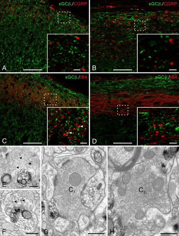
Nitric oxide (NO) has been implicated in pain processing at the spinal level, but the mechanisms mediating its effects remain unclear. In the present work, we studied the organization of the major downstream effector of NO, soluble guanylyl cyclase (sGC), in the superficial dorsal horn of rat. Almost all neurokinin 1 (NK1) receptor-positive neurons in lamina I (a major source of ascending projections) were strongly immunopositive for sGC. Many local circuit neurons in laminae I-II also stained for sGC, but less intensely. Numerous fibers, presumably of unmyelinated primary afferent (C fiber) origin, stained for calcitonin gene-related peptide or isolectin B4, but none of these was immunopositive for sGC. These data, along with immunoelectron microscopy results, imply that unmyelinated primary afferent fibers terminating in the superficial dorsal horn lack sGC. Double labeling showed that neuronal nitric oxide synthase (nNOS) seldom colocalized with sGC, but nNOS-positive structures were frequently closely apposed to sGC-positive structures, suggesting that in the superficial dorsal horn NO acts mainly in a paracrine manner. Our data suggest that the NK1 receptor-positive projection neurons in lamina I are a major target of NO released in superficial dorsal horn. NO may also influence local circuit neurons, but it does not act on unmyelinated primary afferent terminals via sGC.


Figures

Fig. 1
Fig. 1
Western blot analysis of sGCβ1 subunit in rat lumbar spinal cord. The anti-sGCβ1 antibody detected a single band at ∼70 kDa, corresponding to β1 subunit.
Fig. 2
Fig. 2
sGCβ1 immunoperoxidase staining in rat superficial dorsal horn at LM (A-C) and EM (D-F). A, low magnification photomicrograph of immunostaining on transverse section. Staining is seen throughout the superficial dorsal horn, strong in lamina I and weaker in lamina II. B, higher magnification view of the boxed region in A. Somatic staining is strong in lamina I. Numerous large puncta (arrows) and some processes (arrowheads) stain densely in lamina I. In lamina II, weakly sGC-positive somata lie among a dense plexus of stained processes. C, parasagittal sections; stained neurons in lamina I were elongated along the rostro-caudal axis. Immunopositive processes also run rostro-caudally in laminae I and IIo, but more randomly in deeper laminae. D, a positive soma in lamina I (arrowheads pointing to DAB deposits scattered in cytoplasm). A few positive dendrites are also visible (arrows). E, transverse section of a positive dendrite located in lamina I. F, a stained axonal terminal makes synaptic contact onto a dendrite in lamina IIi. Scale bars = 100 μm (A); 20 μm (B, C); 2 μm (D); 0.5 μm (E, F).
Fig. 3
Fig. 3
Double labeling of sGCβ1 with NK1 receptor. Boxed area in A is enlarged in B-D. NK1 receptor-positive somata and dendrites are mainly in lamina I. All NK1 receptor-positive cell bodies contain sGCβ1 staining (arrows). Many of the NK1 receptor-stained processes also stain for sGC (arrowheads). Scale bars = 50 μm (A); 10 μm (B-D).
Fig. 4
Fig. 4
Double labeling of sGCβ1 (green) with GABA (red) in transverse section. A, GABA-stained cell bodies scattered in superficial dorsal horn; the large majority are also stained for sGC. High magnification view of boxed areas in A are shown in B and C. B, three GABA-positive neurons are also immunopositive for sGC (asterisks). C, two sGC-positive cells in lamina II do not express GABA (arrows). Scale bars = 50 μm (A); 10 μm (B and C).
Fig. 5
Fig. 5
The relationship of sGC with primary afferent fibers. A-D: Double labeling of sGCβ1 (green) with two markers for unmyelinated primary afferent fibers, CGRP and IB4 (red) on transverse (A and C) and parasagittal (B and D) sections. Insets are high magnification view of the boxed areas. A and B, CGRP-stained primary afferents terminate mainly in lamina I and IIo. No colocalization of sGC with CGRP was found in transverse or parasagittal sections. C and D, IB4-stained (non-peptidergic) primary afferents terminate mainly in lamina IIi. sGC and IB4 staining are in general unrelated both in transverse and parasagittal section. Occasional contacts are visible (arrowheads), but no clear colocalization. E-H: sGCβ1 immunoperoxidase staining at EM level showing lack of sGC in primary afferent terminals. E, two immunonegative non-glomerular terminals in lamina I that contain dense-core vesicles (arrowheads) close to an immunopositive dendrite. F, an immunonegative terminal (likely to be peptidergic for its content of dense-core vesicles, arrowheads) makes synaptic contact onto an immunopositive dendrite. G, an immunonegative central terminal of type I (C1) synaptic glomerulus in lamina II. H, a negative central terminal of type II (C2) synaptic glomerulus in lamina II. Scale bars = 50 μm (A-D); 5 μm (insets of A-D); 0.5 μm (E-H).
Fig. 6
Fig. 6
The spatial relationship of sGCβ1 and nNOS. nNOS-positive cell bodies are scattered in superficial dorsal horn, most numerous at the ventral border of lamina II. One nNOS-positive cell body is also stained for sGCβ1 (arrow in A). nNOS-positive processes form a dense plexus in lamina IIi, lamina I has moderate nNOS immunoreactivity, and lamina IIo has little. B-D are enlargements of boxed areas in A showing the relationship between the two antigens in different laminae. nNOS-positive puncta are commonly closely apposed to sGC-positive puncta; some of these are circled. Occasional apparent colocalization is also visible (arrowhead). E: Triple labeling with synaptophysin. A few nNOS/sGC contacts (circles) are associated with synaptophysin (arrowheads), while most of those contacts are not. Scale bars = 50 μm (A); 10 μm (B-E).
Fig. 7
Fig. 7
Schematic diagram of the NO-cGMP pathway in superficial dorsal horn. nNOS-positive cells, lying mainly in deep lamina II, produce NO when activated, which diffuses to act on sGC-expressing structures. Concentration of sGC is indicated by darkness of shading. NK1 receptor-expressing projection neurons (black) contain high levels of sGC, and thus may be the primary target of NO. Excitatory and inhibitory interneurons are also potential targets of NO. Unmyelinated primary afferents, which lack sGC, are not affected by NO via the cGMP pathway.

References

    1. Aimar P, Pasti L, Carmignoto G, Merighi A. Nitric oxide-producing islet cells modulate the release of sensory neuropeptides in the rat substantia gelatinosa. J Neurosci. 1998;18:10375–10388. - PMC - PubMed
    1. Bellamy TC, Garthwaite J. The receptor-like properties of nitric oxide-activated soluble guanylyl cyclase in intact cells. Mol Cell Biochem. 2002;230:165–176. - PubMed
    1. Bellamy TC, Wood J, Goodwin DA, Garthwaite J. Rapid desensitization of the nitric oxide receptor, soluble guanylyl cyclase, underlies diversity of cellular cGMP responses. Proc Natl Acad Sci U S A. 2000;97:2928–2933. - PMC - PubMed
    1. Belyantseva IA, Lewin GR. Stability and plasticity of primary afferent projections following nerve regeneration and central degeneration. Eur J Neurosci. 1999;11:457–468. - PubMed
    1. Bernardi PS, Valtschanoff JG, Weinberg RJ, Schmidt HHHW, Rustioni A. Synaptic interactions between primary afferent terminals and GABA and nitric oxide-synthesizing neurons in superficial laminae of the rat spinal cord. J Neurosci. 1995;15:1363–1371. - PMC - PubMed

Publication types

MeSH terms