Comparative Study
. 2006 Feb 28:7:6.
doi: 10.1186/1471-2091-7-6.

Farnesylation or geranylgeranylation? Efficient assays for testing protein prenylation in vitro and in vivo

Affiliations
Comparative Study

Farnesylation or geranylgeranylation? Efficient assays for testing protein prenylation in vitro and in vivo

Wolfgang Benetka et al. BMC Biochem. .

Abstract

Background: Available in vitro and in vivo methods for verifying protein substrates for posttranslational modifications via farnesylation or geranylgeranylation (for example, autoradiography with 3H-labeled anchor precursors) are time consuming (weeks/months), laborious and suffer from low sensitivity.

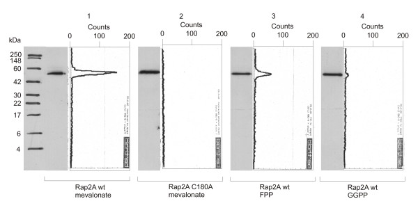
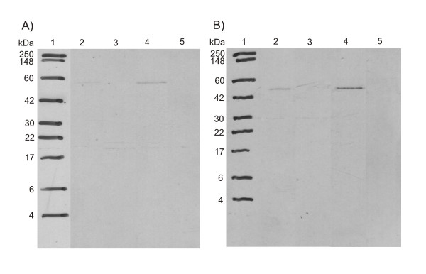
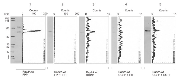
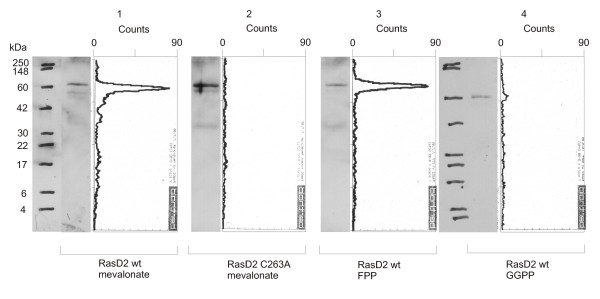
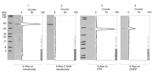
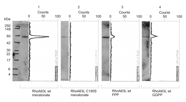
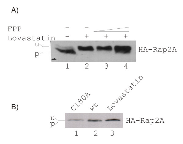
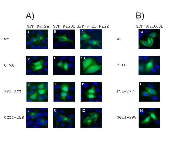
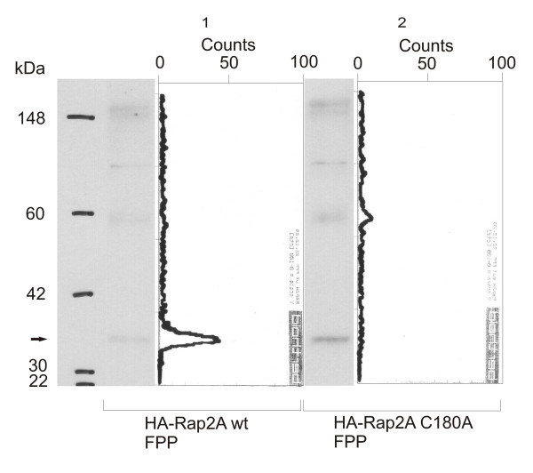
Results: We describe a new technique for detecting prenyl anchors in N-terminally glutathione S-transferase (GST)-labeled constructs of target proteins expressed in vitro in rabbit reticulocyte lysate and incubated with 3H-labeled anchor precursors. Alternatively, hemagglutinin (HA)-labeled constructs expressed in vivo (in cell culture) can be used. For registration of the radioactive marker, we propose to use a thin layer chromatography (TLC) analyzer. As a control, the protein yield is tested by Western blotting with anti-GST- (or anti-HA-) antibodies on the same membrane that has been previously used for TLC-scanning. These protocols have been tested with Rap2A, v-Ki-Ras2 and RhoA (variant RhoA63L) including the necessary controls. We show directly that RasD2 is a farnesylation target.

Conclusion: Savings in time for experimentation and the higher sensitivity for detecting 3H-labeled lipid anchors recommend the TLC-scanning method with purified GST- (or HA-) tagged target proteins as the method of choice for analyzing their prenylation capabilities in vitro and in vivo and, possibly, also for studying the myristoyl and palmitoyl posttranslational modifications.


Figures

Figure 1
Figure 1
Western blots and TLC scanning results for Rap2A with radioactive prenyl anchor precursors. Western Blot and corresponding scans from TLC linear analyzer of wildtype GST-Rap2A-fusion protein translated with [3H]mevalonic acid (lane 1), GST-Rap2A C180A with [3H]mevalonic acid (lane 2), GST-Rap2A with [3H]FPP (lane 3) and GST-Rap2A with [3H]GGPP (lane 4). There is significant incorporation of a product of mevalonic acid (lane 1) as well as FPP (lane 3), while incorporation of GGPP is close to the detection limit (lane 4), suggesting that Rap2A is primarily a farnesylation target.
Figure 2
Figure 2
Autoradiographs of Rap2A after exposure to radioactive prenyl anchor precursors. Fluorography of GST-Rap2A-fusion protein on a Western membrane after treatment with En3Hance-spray (2-methyl-naphtalene, Perkin-Elmer), showing a protein size marker in lane 1, wildtype GST-Rap2A translated with [3H]mevalonic acid in lane 2, GST-Rap2A C180A with [3H]mevalonic acid in lane 3, GST-Rap2A with [3H]FPP in lane 4 and GST-Rap2A with [3H]GGPP in lane 5. A) film after exposure for 7 days, B) film after exposure for 20 days at -80°C. There is no sign of incorporation of GGPP as detected with the TLC-scanner, underscoring the higher sensitivity of our new method. It should be noted that it is difficult to evenly spread the En3Hance-substance over all membrane area. Therefore, it is not surprising that the relative signal intensities are not identical between TLC scanning and autoradiography.
Figure 3
Figure 3
Western blots and TLC scanning results for Rap2A incubated with prenyltransferase inhibitors. Western Blot and corresponding scans from TLC linear analyzer of wildtype GST-Rap2A-fusion protein translated with [3H]FPP (lane 1), with [3H]FPP and 50 μM FTI-277 (lane 2), with [3H]GGPP (lane 3), with [3H]GGPP and 50 μM FTI-277 (lane 4), and with [3H]GGPP and 50 μM GGTI-298 (lane 5). There is no incorporation of FPP with FTI (lane 2), and there is also no incorporation of GGPP with FTI (lane 4), while a hardly detectable signal remains with GGTI (lane 5), suggesting that Rap2A is recognized only by farnesyltransferase. However, the enzyme shows some cross-reactivity with GGPP.
Figure 4
Figure 4
Western blots and TLC scanning results for RasD2 with radioactive prenyl anchor precursors. Western Blot and corresponding scans from TLC linear analyzer of wildtype GST-RasD2-fusion protein translated with [3H]mevalonic acid (lane 1), GST-RasD2 C263A with [3H]mevalonic acid (lane 2), GST-RasD2 with [3H]FPP (lane 3) and GST-RasD2 with [3H]GGPP (lane 4). There is significant incorporation of a product of mevalonic acid (lane 1) as well as FPP (lane 3), while incorporation of GGPP is close to the detection limit (lane 4), suggesting that RasD2 is recognized primarily by the FTase.
Figure 5
Figure 5
Western blots and TLC scanning results for v-Ki-Ras2 (K-Ras-4B) with radioactive prenyl anchor precursors. Western Blot and corresponding scans from TLC linear analyzer of wildtype GST-v-Ki-Ras2-fusion protein translated with [3H]mevalonic acid (lane 1), GST- v-Ki-Ras2 C185A with [3H]mevalonic acid (lane 2), GST-K-Ras with [3H]FPP (lane 3) and GST-K-Ras with [3H]GGPP (lane 4). There is incorporation of a product of mevalonic acid (lane 1) and FPP (lane 3) and also a reduced but noticeable amount of GGPP (lane 4), supporting the view of alternative geranylgeranylation of K-Ras in the absence of farnesylation.
Figure 6
Figure 6
Western blots and TLC scanning results for RhoA63L with radioactive prenyl anchor precursors. Western Blot and corresponding scans from TLC linear analyzer of wildtype GST-RhoA63L-fusion protein translated with [3H]mevalonic acid (lane 1), GST-RhoA63L mutant C190S with [3H]mevalonic acid (lane 2), GST-RhoA63L with [3H]FPP (lane 3) and GST-RhoA63L with [3H]GGPP (lane 4). There is significant incorporation of a product of mevalonic acid (lane 1) as well as of GGPP (lane 4). The signal for FPP attachment is reduced, although more protein is detected (lane 3). This confirms GGPP as preferred substrate.
Figure 7
Figure 7
Mobility changes of the prenylated protein form: Immunoblot analysis of Rap2A. Western blot analysis has been performed on lysates of exponentially growing cells. U denotes the unmodified, P the prenylated form of Rap2A. A) HeLa cells have been transiently transfected with HA-Rap2A (lanes 1–4). Upon treatment with lovastatin (lane 2), the signal representing prenylated (p) Rap2A disappeared. This effect could be reversed by adding FPP (lanes 3 and 4), but not by adding GGPP (data not shown). B) HeLa cells have been transiently transfected with HA-Rap2A (lanes 2 and 3) or HA-Rap2A with a cysteine-to-alanine mutation within the C-terminal CAAX prenylation motif (lane 1, mutation C180A). The mutation and also the lovastatin treatment prevent the prenylation of HA-Rap2A.
Figure 8
Figure 8
Localization of N-terminal GFP-constructs of Rap2A, RasD2, v-Ki-Ras2 and RhoA63L in HeLa cells. HeLa cells were analysed by fluorescence microscopy after transfection with the following constructs: inserts 1, 3 and 4 – GFP-Rap2A; insert 2 – GFP-Rap2A C180A; inserts 5, 7 and 8 – GFP-RasD2; insert 6 – GFP-RasD2 C263A; inserts 9, 11 and 12 –GFP-v-Ki-Ras2; insert 10 – GFP-v-Ki-Ras2 C185A; inserts 13, 15 and 16 – GFP-RhoA63L; insert 14 – GFP-RhoA63L C190S. Nuclei were co-stained with DAPI (blue color). A) GFP-Rap2A, GFP-RasD2 and GFP-v-Ki-Ras2 are membrane-localized with (4, 8, 12) or without (1, 5, 9) GGTI-298 treatment. Mutation of the Cys in the CaaX box (2, 6, 10) or treatment with FTI-277 (3, 7, 11) cause mislocalization and accumulation of the fusion proteins in the nucleus. B) GFP-RhoA is membrane localized with (15) or without (13) FTI-277 treatment. Mutation of the Cys in the CaaX box (14) or treatment with GGTI-298 (16) cause mislocalization and accumulation of RhoA in the nucleus.
Figure 9
Figure 9
Western blots and TLC scanning results for Rap2A with radioactive FPP in vivo. Western Blot and corresponding scans from TLC linear analyzer of wild-type HA-Rap2A-fusion protein (lane 1) and HA-Rap2A C180A (lane 2) immunoprecipitated from HeLa-cells, treated with lovastatin after transfection with the respective plasmid construct and exposed to 3H-FPP. There is significant incorporation of FPP into the wild-type protein (lane 1), but no signal is detected for the C180A mutant (lane 2), demonstrating the applicability of the approach for in vivo labeling experiments.

Similar articles

Cited by

References

    1. Glomset JA, Gelb MH, Farnsworth CC. Prenyl proteins in eukaryotic cells: a new type of membrane anchor. Trends Biochem Sci. 1990;15:139–142. doi: 10.1016/0968-0004(90)90213-U. - DOI - PubMed
    1. Moores SL, Schaber MD, Mosser SD, Rands E, O'Hara MB, Garsky VM, Marshall MS, Pompliano DL, Gibbs JB. Sequence dependence of protein isoprenylation. J Biol Chem. 1991;266:14603–14610. - PubMed
    1. Caplin BE, Hettich LA, Marshall MS. Substrate characterization of the Saccharomyces cerevisiae protein farnesyltransferase and type-I protein geranylgeranyltransferase. Biochim Biophys Acta. 1994;1205:39–48. - PubMed
    1. Zhang FL, Casey PJ. Protein prenylation: molecular mechanisms and functional consequences. Annu Rev Biochem. 1996;65:241–269. doi: 10.1146/annurev.bi.65.070196.001325. - DOI - PubMed
    1. Maurer-Stroh S, Washietl S, Eisenhaber F. Protein prenyltransferases. Genome Biol. 2003;4:212. doi: 10.1186/gb-2003-4-4-212. - DOI - PMC - PubMed

Publication types