

Ultrasound biomicroscopy findings of 25 G Transconjuctival Sutureless (TSV) and conventional (20G) pars plana sclerotomy in the same patient
- PMID: 16507105
- PMCID: PMC1403799
- DOI: 10.1186/1471-2415-6-7
Ultrasound biomicroscopy findings of 25 G Transconjuctival Sutureless (TSV) and conventional (20G) pars plana sclerotomy in the same patient
Abstract
Background: Transconjunctival Sutureless Vitrectomy (TSV) is a recent advancement in vitreo-retinal surgical techniques involving the use of 25 G instruments through self-sealing sclerotomies. It has been hypothesized that there may be less chance of vitreous and retinal herniation in the scleral wound as compared to conventional sclerotomy incision. However there are no reports on differences in 20 gauge and 25 gauge sclerotomies using ultrasound biomicroscopy (UBM). We report herein the differences in sclerotomies undertaken with 20 gauge (G) and 25 gauge instruments in the same patient.
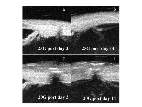
Case presentation: Ultrasound biomicroscopy of the sclerotomy sites was done in the same patient in whom both 20 G and 25 G sclerotomies had to be constructed during pars plana vitrectomy and the differences were studied. On day 2, we observed a wide gape at the site that had been enlarged using a 20G MVR blade. In contrast, the other two sites made transconjunctivally using the 25G trocar showed only a mild gape. Significant gape continued to persist at the subsequent evaluations on day 7 and day 14 only at the port, which had been enlarged.
Conclusion: Healing of a 25 G sclerotomy is expectedly quite rapid, with inability to detect the site of sclerotomy in a short duration of 2 weeks post-operatively. This is as opposed to conventional sclerotomies, which might take up to 6-8 weeks post-operatively for complete opposition.
Figures
Similar articles
-
Transconjunctival sutureless 25-gauge versus 20-gauge standard vitrectomy: correlation between corneal topography and ultrasound biomicroscopy measurements of sclerotomy sites.Cornea. 2010 Jan;29(1):19-25. doi: 10.1097/ICO.0b013e3181ab98ae. Cornea. 2010. PMID: 19907299 Clinical Trial.
-
Ultrasound biomicroscopy in recently postoperative 23-gauge transconjunctival vitrectomy sutureless self-sealing sclerotomy.Retina. 2009 Oct;29(9):1305-9. doi: 10.1097/IAE.0b013e3181b09487. Retina. 2009. PMID: 19696703 Clinical Trial.
-
Ultrasound biomicroscopy of sclerotomy sites after pars plana vitrectomy for diabetic vitreous hemorrhage.Ophthalmology. 2000 Sep;107(9):1729-36. doi: 10.1016/s0161-6420(00)00213-x. Ophthalmology. 2000. PMID: 10964837
-
Transconjunctival 20-gauge pars plana vitrectomy using a single entry cannulated sutureless system.Retina. 2009 Oct;29(9):1294-8. doi: 10.1097/IAE.0b013e3181aa8e3b. Retina. 2009. PMID: 19696700
-
[Dry transconjunctival sutureless 25-gauge vitrectomy in the treatment of pediatric cataract].Zhonghua Yan Ke Za Zhi. 2009 Aug;45(8):762-5. Zhonghua Yan Ke Za Zhi. 2009. PMID: 20021895 Review. Chinese.
Cited by
-
Efficacy and Safety of Intravitreal Conbercept, Ranibizumab, and Triamcinolone on 23-Gauge Vitrectomy for Patients with Proliferative Diabetic Retinopathy.J Ophthalmol. 2018 Jun 25;2018:4927259. doi: 10.1155/2018/4927259. eCollection 2018. J Ophthalmol. 2018. PMID: 30046459 Free PMC article.
-
Modified incision in 25-gauge vitrectomy in the creation of a tunneled airtight sclerotomy: an ultrabiomicroscopic study.Graefes Arch Clin Exp Ophthalmol. 2007 Sep;245(9):1281-8. doi: 10.1007/s00417-006-0533-x. Epub 2007 Feb 21. Graefes Arch Clin Exp Ophthalmol. 2007. PMID: 17318571
-
Sutureless vitrectomy.Indian J Ophthalmol. 2008 Nov-Dec;56(6):453-8. doi: 10.4103/0301-4738.43364. Indian J Ophthalmol. 2008. PMID: 18974514 Free PMC article. Review.
-
23-gauge pars plana vitrectomy for management of posteriorly dislocated crystalline lens.Clin Ophthalmol. 2011;5:1737-43. doi: 10.2147/OPTH.S22331. Epub 2011 Dec 8. Clin Ophthalmol. 2011. PMID: 22205834 Free PMC article.
-
Surgical outcomes of 25-gauge pars plana vitrectomy for diabetic tractional retinal detachment.Eye (Lond). 2015 Sep;29(9):1213-9. doi: 10.1038/eye.2015.126. Epub 2015 Jul 17. Eye (Lond). 2015. PMID: 26183284 Free PMC article.
References
-
- Boker T, Spitznas M. Ultrasound biomicroscopy for examination of the sclerotomy sites after parsplana vitrectomy. Am J Ophthalmol. 1994;118:813–815. - PubMed
-
- Bhende M, Agraharam SG, Gopal L, Sumasri K, Sukumar B, George J, Sharma T, Shanmugam MP, Bhende PS, Shetty NS, Agarwal RN, Despande DA. Ultrasound biomicroscopy of sclerotomy sites after pars plana vitrectomy for diabetic vitreous hemorrhage. Ophthalmology. 2000;107:1729–36. doi: 10.1016/S0161-6420(00)00213-X. - DOI - PubMed
Publication types
MeSH terms
LinkOut - more resources
Full Text Sources
Miscellaneous
