

Perlecan, a candidate gene for the CAPB locus, regulates prostate cancer cell growth via the Sonic Hedgehog pathway
- PMID: 16507112
- PMCID: PMC1421430
- DOI: 10.1186/1476-4598-5-9
Perlecan, a candidate gene for the CAPB locus, regulates prostate cancer cell growth via the Sonic Hedgehog pathway
Abstract
Background: Genetic studies associated the CAPB locus with familial risk of brain and prostate cancers. We have identified HSPG2 (Perlecan) as a candidate gene for CAPB. Previously we have linked Perlecan to Hedgehog signaling in Drosophila. More recently, we have demonstrated the importance of Hedgehog signaling in humans for advanced prostate cancer.
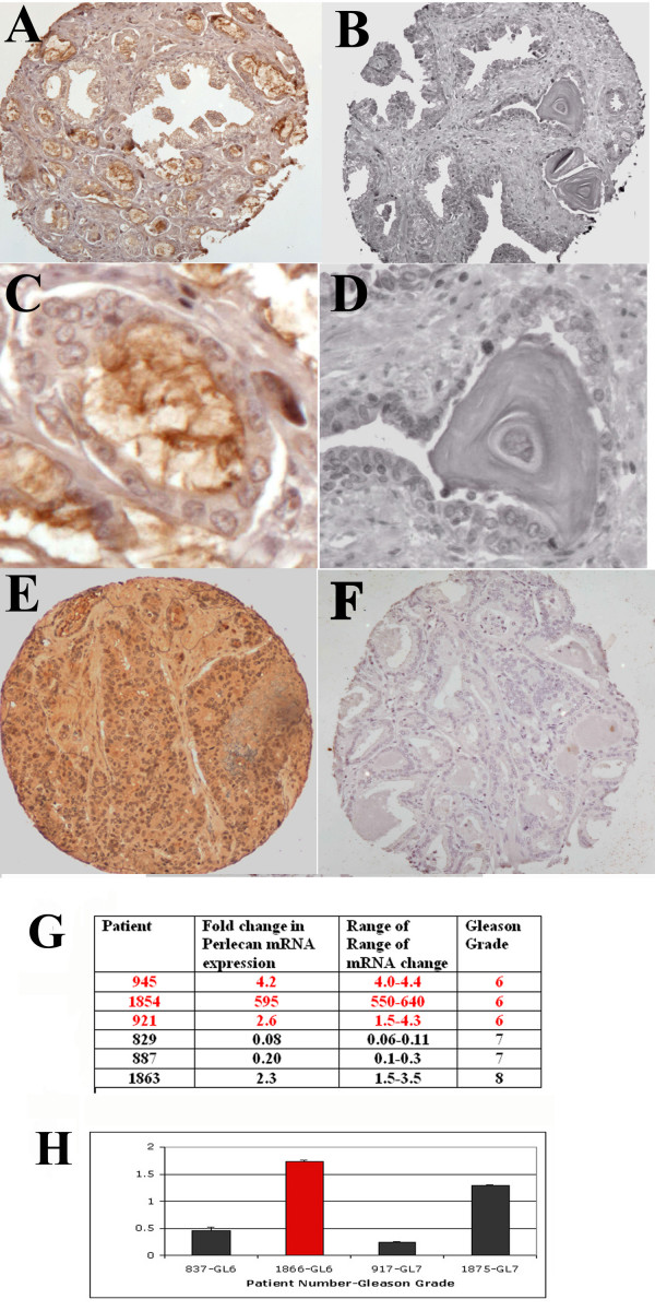
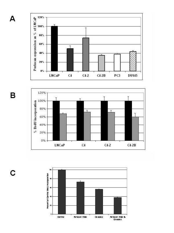
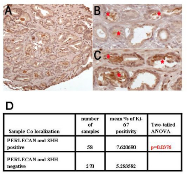
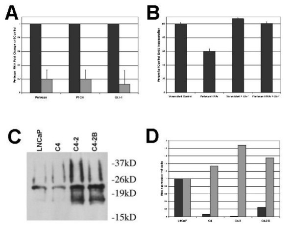
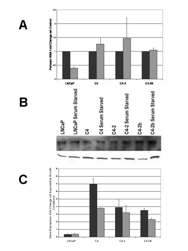
Results: Here we demonstrate Perlecan expression in prostate cancer, and its function in prostate cancer cell growth through interaction and modulation of Sonic Hedgehog (SHH) signaling. Perlecan expression in prostate cancer tissues correlates with a high Gleason score and rapid cell proliferation. Perlecan is highly expressed in prostate cancer cell lines, including androgen insensitive cell lines and cell lines selected for metastatic properties. Inhibition of Perlecan expression in these cell lines decreases cell growth. Simultaneous blockade of Perlecan expression and androgen signaling in the androgen-sensitive cell line LNCaP was additive, indicating the independence of these two pathways. Perlecan expression correlates with SHH in tumor tissue microarrays and increased tumor cell proliferation based on Ki-67 immunohistochemistry. Inhibition of Perlecan expression by siRNA in prostate cancer cell lines decreases SHH signaling while expression of the downstream SHH effector GLI1 rescues the proliferation defect. Perlecan forms complexes with increasing amounts of SHH that correlate with increasing metastatic potential of the prostate cancer cell line. SHH signaling also increases in the more metastatic cell lines. Metastatic prostate cancer cell lines grown under serum-starved conditions (low androgen and growth factors) resulted in maintenance of Perlecan expression. Under low androgen, low growth factor conditions, Perlecan expression level correlates with the ability of the cells to maintain SHH signaling.
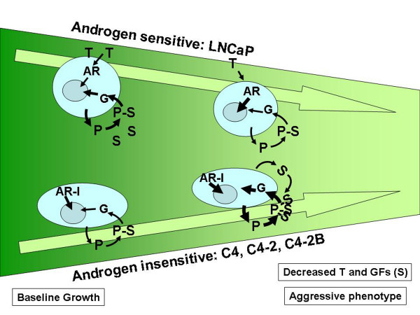
Conclusion: We have demonstrated that Perlecan, a candidate gene for the CAPB locus, is a new component of the SHH pathway in prostate tumors and works independently of androgen signaling. In metastatic tumor cells increased SHH signaling correlates with the maintenance of Perlecan expression and more Perlecan-SHH complexes. Perlecan is a proteoglycan that regulates extracellular and stromal accessibility to growth factors such as SHH, thus allowing for the maintenance of SHH signaling under growth factor limiting conditions. This proteoglycan represents an important central regulator of SHH activity and presents an ideal drug target for blocking SHH effects.
Figures
Similar articles
-
Perlecan signaling: helping hedgehog stimulate prostate cancer growth.Int J Biochem Cell Biol. 2006;38(11):1855-61. doi: 10.1016/j.biocel.2006.03.022. Epub 2006 Apr 25. Int J Biochem Cell Biol. 2006. PMID: 16750652 Review.
-
Inhibition of prostate cancer proliferation by interference with SONIC HEDGEHOG-GLI1 signaling.Proc Natl Acad Sci U S A. 2004 Aug 24;101(34):12561-6. doi: 10.1073/pnas.0404956101. Epub 2004 Aug 16. Proc Natl Acad Sci U S A. 2004. PMID: 15314219 Free PMC article.
-
Sonic hedgehog and androgen signaling in tumor and stromal compartments drives epithelial-mesenchymal transition in prostate cancer.Scand J Urol. 2014 Dec;48(6):523-32. doi: 10.3109/21681805.2014.898336. Epub 2014 Mar 19. Scand J Urol. 2014. PMID: 25356787
-
Carnosol inhibits Hedgehog signaling pathway in both LNCaP and DU145 prostate cancer cell lines.Cell Mol Biol (Noisy-le-grand). 2017 Aug 30;63(8):104-108. doi: 10.14715/cmb/2017.63.8.22. Cell Mol Biol (Noisy-le-grand). 2017. PMID: 28886322
-
Sonic Hedgehog signaling in advanced prostate cancer.Cell Mol Life Sci. 2006 Feb;63(4):435-48. doi: 10.1007/s00018-005-5389-4. Cell Mol Life Sci. 2006. PMID: 16389455 Free PMC article. Review.
Cited by
-
Proteoglycans and their roles in brain cancer.FEBS J. 2013 May;280(10):2399-417. doi: 10.1111/febs.12109. Epub 2013 Feb 6. FEBS J. 2013. PMID: 23281850 Free PMC article. Review.
-
Microfabricated electrospun collagen membranes for 3-D cancer models and drug screening applications.Biomacromolecules. 2009 Aug 10;10(8):2019-32. doi: 10.1021/bm8012764. Biomacromolecules. 2009. PMID: 19624098 Free PMC article.
-
A central function for perlecan in skeletal muscle and cardiovascular development.J Cell Biol. 2008 Apr 21;181(2):381-94. doi: 10.1083/jcb.200708022. J Cell Biol. 2008. PMID: 18426981 Free PMC article.
-
Matrilysin/matrix metalloproteinase-7(MMP7) cleavage of perlecan/HSPG2 creates a molecular switch to alter prostate cancer cell behavior.Matrix Biol. 2014 Jun;36:64-76. doi: 10.1016/j.matbio.2014.04.005. Epub 2014 May 14. Matrix Biol. 2014. PMID: 24833109 Free PMC article.
-
Heparanase expression and activity influences chondrogenic and osteogenic processes during endochondral bone formation.Bone. 2008 Oct;43(4):689-99. doi: 10.1016/j.bone.2008.05.022. Epub 2008 Jun 6. Bone. 2008. PMID: 18589009 Free PMC article.
References
-
- Conlon EM, Goode EL, Gibbs M, Stanford JL, Badzioch M, Janer M, Kolb S, Hood L, Ostrander EA, Jarvik GP, Wijsman EM. Oligogenic segregation analysis of hereditary prostate cancer pedigrees: evidence for multiple loci affecting age at onset. Int J Cancer. 2003;105:630–635. doi: 10.1002/ijc.11128. - DOI - PubMed
-
- Gibbs M, Stanford JL, McIndoe RA, Jarvik GP, Kolb S, Goode EL, Chakrabarti L, Schuster EF, Buckley VA, Miller EL, Brandzel S, Li S, Hood L, Ostrander EA. Evidence for a rare prostate cancer-susceptibility locus at chromosome 1p36. Am J Hum Genet. 1999;64:776–787. doi: 10.1086/302287. - DOI - PMC - PubMed
Publication types
MeSH terms
Substances
Grants and funding
LinkOut - more resources
Full Text Sources
Other Literature Sources
Medical
