

Long term evaluation of disease progression through the quantitative magnetic resonance imaging of symptomatic knee osteoarthritis patients: correlation with clinical symptoms and radiographic changes
- PMID: 16507119
- PMCID: PMC1526551
- DOI: 10.1186/ar1875
Long term evaluation of disease progression through the quantitative magnetic resonance imaging of symptomatic knee osteoarthritis patients: correlation with clinical symptoms and radiographic changes
Abstract
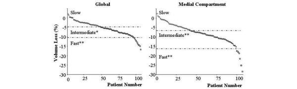
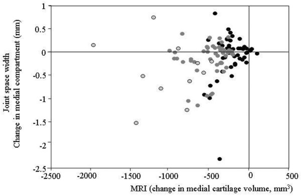
The objective of this study was to further explore the cartilage volume changes in knee osteoarthritis (OA) over time using quantitative magnetic resonance imaging (qMRI). These were correlated with demographic, clinical, and radiological data to better identify the disease risk features. We selected 107 patients from a large trial (n = 1,232) evaluating the effect of a bisphosphonate on OA knees. The MRI acquisitions of the knee were done at baseline, 12, and 24 months. Cartilage volume from the global, medial, and lateral compartments was quantified. The changes were contrasted with clinical data and other MRI anatomical features. Knee OA cartilage volume losses were statistically significant compared to baseline values: -3.7 +/- 3.0% for global cartilage and -5.5 +/- 4.3% for the medial compartment at 12 months, and -5.7 +/- 4.4% and -8.3 +/- 6.5%, respectively, at 24 months. Three different populations were identified according to cartilage volume loss: fast (n = 11; -13.2%), intermediate (n = 48; -7.2%), and slow (n = 48; -2.3%) progressors. The predictors of fast progressors were the presence of severe meniscal extrusion (p = 0.001), severe medial tear (p = 0.005), medial and/or lateral bone edema (p = 0.03), high body mass index (p < 0.05, fast versus slow), weight (p < 0.05, fast versus slow) and age (p < 0.05 fast versus slow). The loss of cartilage volume was also slightly associated with less knee pain. No association was found with other Western Ontario McMaster Osteoarthritis Index (WOMAC) scores, joint space width, or urine biomarker levels. Meniscal damage and bone edema are closely associated with more cartilage volume loss. These data confirm the significant advantage of qMRI for reliably measuring knee structural changes at as early as 12 months, and for identifying risk factors associated with OA progression.
Figures
References
-
- Bingham C, Cline G, Cohen G, Wenderoth D, Conaghan P, Buckland-Wright C, Beary J, Dougados M, Strand V, Meyer J. Predictors of structural progression in knee osteoarthritis over 24 Months [abstract] Arthritis Rheum. 2004;50(supp 9):254.
-
- Raynauld JP, Kauffmann C, Beaudoin G, Berthiaume MJ, de Guise JA, Bloch DA, Camacho F, Godbout B, Altman RD, Hochberg M, et al. Reliability of a quantification imaging system using magnetic resonance images to measure cartilage thickness and volume in human normal and osteoarthritic knees. Osteoarthritis Cartilage. 2003;11:351–360. doi: 10.1016/S1063-4584(03)00029-3. - DOI - PubMed
Publication types
MeSH terms
Substances
LinkOut - more resources
Full Text Sources
Other Literature Sources
Medical
