. 2006;8(1):R23.
doi: 10.1186/ar1877. Epub 2006 Jan 3.

Fibroblast activation protein alpha is expressed by chondrocytes following a pro-inflammatory stimulus and is elevated in osteoarthritis

Affiliations

Fibroblast activation protein alpha is expressed by chondrocytes following a pro-inflammatory stimulus and is elevated in osteoarthritis

Jennifer M Milner et al. Arthritis Res Ther. 2006.

Abstract

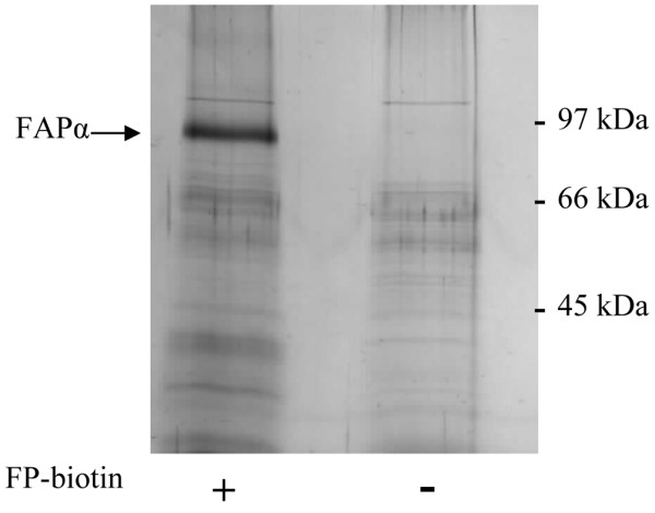
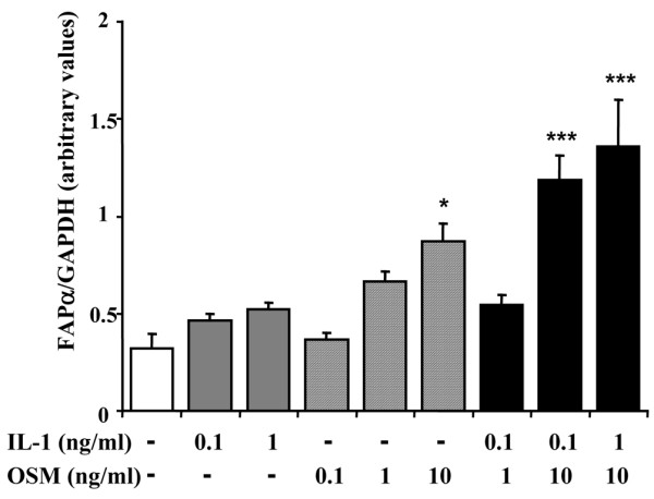
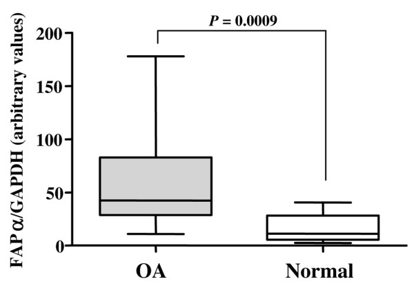
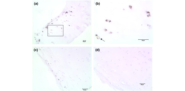
Arthritis is characterised by the proteolytic degradation of articular cartilage leading to a loss of joint function. Articular cartilage is composed of an extracellular matrix of proteoglycans and collagens. We have previously shown that serine proteinases are involved in the activation cascades leading to cartilage collagen degradation. The aim of this study was to use an active-site probe, biotinylated fluorophosphonate, to identify active serine proteinases present on the chondrocyte membrane after stimulation with the pro-inflammatory cytokines IL-1 and oncostatin M (OSM), agents that promote cartilage resorption. Fibroblast activation protein alpha (FAPalpha), a type II integral membrane serine proteinase, was identified on chondrocyte membranes stimulated with IL-1 and OSM. Real-time PCR analysis shows that FAPalpha gene expression is up-regulated by this cytokine combination in both isolated chondrocytes and cartilage explant cultures and is significantly higher in cartilage from OA patients compared to phenotypically normal articular cartilage. Immunohistochemistry analysis shows FAPalpha expression on chondrocytes in the superficial zone of OA cartilage tissues. This is the first report demonstrating the expression of active FAPalpha on the chondrocyte membrane and elevated levels in cartilage from OA patients. Its cell surface location and expression profile suggest that it may have an important pathological role in the cartilage turnover prevalent in arthritic diseases.


Figures

Figure 1
Figure 1
Biotinylated fluorophosphonate (FP-biotin) labelling of fibroblast activation protein alpha (FAPα) on chondrocyte membranes. Membrane extracts isolated from IL-1 plus oncostatin M stimulated chondrocytes were treated with or without FP-biotin. Labelled proteinases were isolated using streptavidin-agarose beads and eluted with reducing SDS-PAGE loading buffer. Proteins were separated by SDS-PAGE and stained with colloidal Coomassie. The 97 kDa protein was identified by mass spectrometry to be FAPα.
Figure 2
Figure 2
FAPα gene expression is upregulated by IL-1 and oncostatin M (OSM) in chondrocytes. SW1353 cells were treated with combinations of IL-1 and OSM for 24 h. Total RNA was extracted and FAPα gene expression determined by real-time PCR as described in Materials and methods. The data are presented relative to GAPDH, and are representative of four separate experiments. * = P < 0.05, *** = P < 0.001 versus control
Figure 3
Figure 3
FAPα gene expression is induced in resorbing cartilage. Bovine nasal cartilage chips were cultured in medium with or without IL-1 (1 ng/ml) and oncostatin M (OSM; 10 ng/ml) for 14 days. At day 7, medium was removed and the cartilage was replenished with identical reagents. Cartilage and medium were harvested at days 0, 1, 3, 5, 7, 8, 10, 12 and 14. Each time-point and condition were performed in triplicate. As a measure of collagen, the levels of hydroxyproline released into the media from unstimulated (control) and IL-1/OSM stimulated cartilage were assayed and cumulative hydroxyproline release is shown. Values are the mean ± standard error of the mean. RNA was extracted from cartilage and FAPα gene expression was determined by real-time PCR as described in Materials and methods. The data are presented relative to 18S and show fold induction of FAPα by IL-1/OSM compared to control treatments.
Figure 4
Figure 4
FAPα gene expression is upregulated in osteoarthritic cartilage. Total RNA was extracted from osteoarthritic hip cartilage (n = 14) and phenotypically normal hip cartilage from patients with femoral neck fracture (n = 12). FAPα gene expression was determined by real-time PCR as described in Materials and methods. The data are presented relative to GAPDH. FAPα gene expression is significantly higher in osteoarthritic cartilage (OA) compared to normal cartilage (P = 0.0009).
Figure 5
Figure 5
Immunolocalisation of FAPα protein in osteoarthritic (OA) cartilage. (a) FAPα in OA cartilage specimen 1. Note positive staining (brown/black) of cells in the superficial zone. Boxed region represents low-power view of Figure 5b. (b) High-power view of FAPα in OA cartilage specimen 1. Note positive staining of the chondrocyte membrane (arrow). (c) FAPα in OA cartilage specimen 2. Note positive staining of cells in the superficial zone. (d) OA cartilage specimen 2 treated with non-immune mouse IgG as a negative control for FAPα.

References

    1. Goldring MB. The role of the chondrocyte in osteoarthritis. Arthritis Rheum. 2000;43:1916–1926. doi: 10.1002/1529-0131(200009)43:9<1916::AID-ANR2>3.0.CO;2-I. - DOI - PubMed
    1. Birkedal-Hansen H, Moore WG, Bodden MK, Windsor LJ, Birkedal-Hansen B, DeCarlo A, Engler JA. Matrix metalloproteinases: a review. Crit Rev Oral Biol Med. 1993;4:197–250. - PubMed
    1. Hollander AP, Pidoux I, Reiner A, Rorabeck C, Bourne R, Poole AR. Damage to type II collagen in aging and osteoarthritis starts at the articular surface, originates around chondrocytes, and extends into the cartilage with progressive degeneration. J Clin Invest. 1995;96:2859–2869. - PMC - PubMed
    1. Clark IM, Parker AE. Metalloproteinases: their role in arthritis and potential as therapeutic targets. Expert Opin Ther Targets. 2003;7:19–34. doi: 10.1517/14728222.7.1.19. - DOI - PubMed
    1. Kevorkian L, Young DA, Darrah C, Donell ST, Shepstone L, Porter S, Brockbank SM, Edwards DR, Parker AE, Clark IM. Expression profiling of metalloproteinases and their inhibitors in cartilage. Arthritis Rheum. 2004;50:131–141. doi: 10.1002/art.11433. - DOI - PubMed

Publication types

MeSH terms