. 2006;7(2):R12.
doi: 10.1186/gb-2006-7-2-r12. Epub 2006 Feb 20.

Lineage-specific expansion of proteins exported to erythrocytes in malaria parasites

Affiliations

Lineage-specific expansion of proteins exported to erythrocytes in malaria parasites

Tobias J Sargeant et al. Genome Biol. 2006.

Abstract

Background: The apicomplexan parasite Plasmodium falciparum causes the most severe form of malaria in humans. After invasion into erythrocytes, asexual parasite stages drastically alter their host cell and export remodeling and virulence proteins. Previously, we have reported identification and functional analysis of a short motif necessary for export of proteins out of the parasite and into the red blood cell.

Results: We have developed software for the prediction of exported proteins in the genus Plasmodium, and identified exported proteins conserved between malaria parasites infecting rodents and the two major causes of human malaria, P. falciparum and P. vivax. This conserved 'exportome' is confined to a few subtelomeric chromosomal regions in P. falciparum and the synteny of these and surrounding regions is conserved in P. vivax. We have identified a novel gene family PHIST (for Plasmodium helical interspersed subtelomeric family) that shares a unique domain with 72 paralogs in P. falciparum and 39 in P. vivax; however, there is only one member in each of the three species studied from the P. berghei lineage.

Conclusion: These data suggest radiation of genes encoding remodeling and virulence factors from a small number of loci in a common Plasmodium ancestor, and imply a closer phylogenetic relationship between the P. vivax and P. falciparum lineages than previously believed. The presence of a conserved 'exportome' in the genus Plasmodium has important implications for our understanding of both common mechanisms and species-specific differences in host-parasite interactions, and may be crucial in developing novel antimalarial drugs to this infectious disease.


Figures
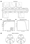
Figure 1
Figure 1
ExportPred: Architecture and performance. (a) The architecture of the ExportPred GHMM. The GHMM progresses from left to right, beginning with an amino-terminal methionine and terminating at a stop codon. Length probability densities are shown for non-geometric states. Tail states and the KLD spacer state are modelled by geometric distributions. (b) ROC curves for the ExportPred model comparing the training against the five described negative sets. (c) False discovery rate as a function of score threshold, calculated using the training set and the P.f negative set, and assuming 10% of the P. falciparum proteome is exported. (d) Comparison of predictions made by ExportPred using the default threshold of 4.3 with those published in [6,7]. The -rifin set is exclusive of any sequence annotated as rifin or stevor, whereas the +rifin set includes these sequences.
Figure 2
Figure 2
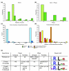
ExportPred: Training sets and validation. (a) Boxplots of scores of two positive sequence sets and five negative sequence sets. The chosen score threshold of 4.3 is marked. Both positive sets are well separated from all negative sets. Poorly scoring outliers in the postive sets can largely be ascribed to incorrect gene models and Rif and Stevor pseudogenes. (b) Two-dimensional plot of P. falciparum proteins decomposed by scores of the ExportPred states for the PEXEL motif and for the signal sequence. Small black dots indicate proteins with full model scores <4.3 and blue dots with scores ≥ 4.3. The three positive and four negative GFP fusions described are marked with green and red dots, respectively, and the nine yellow dots are, from left to right, RESA, HRPIII, KAHRP, PFA0475w (Rifin), R45, MESA, PfEMP3, PFC0025c (Stevor), and GBP130. (c) Experimental verification of a number of ExportPred predictions above (green) and below (red) the chosen threshold. GFP fusions to three positive predictions (PFI1780w, PFE0055c, PFI1755c) are exported successfully into the red blood cell cytosol. Fusion proteins to three negative predictions (PFE0360c, PF14_0607, PFE0355w) accumulate in the parasitophorous vacuole, indicating a functional signal sequence but no functional export motif. One GFP fusion (PF10_0321) appears to be targeted to the mitochondrion. ExportPred scores are indicated in parentheses.
Figure 3
Figure 3
Plasmodium 'exportome' statistics. (a) Distribution of exon counts in genes with PEXEL export signatures compared with all P. falciparum genes, demonstrating a clear trend towards two exon genes in the P. falciparum exportome. (b) First intron phase for PEXEL exported genes compared with all P. falciparum genes, showing an extremely strong trend towards a phase 0 first intron amongst genes with export signatures. (c) Counts of classic (intron between signal sequence and PEXEL) and non-classic genes in the P. falciparum exportome, stratified by exon count. (d) Distributions of the number of predicted transmembrane domains for exported P. falciparum proteins, Rifins and Stevors, and the P. falciparum proteome as a whole. Rifins and Stevors are, in general, predicted to have two transmembrane domains, and members of the remaining complement of the P. falciparum exportome are slightly less likely to be soluble than P. falciparum proteins in general, and are also less likely to be multi-membrane spanning. (e) Comparison of the P. falciparum exportome with hybrid exportomes of the P. vivax and P. berghei lineages. Numbers of PEXEL exported uniques and families are shown, as well as any previously described families and uniques not apparently exported by PEXEL mediated mechanisms. Web logos constructed from instances of the motif in the three exportomes are also shown. References to gene families from species other than P. falciparum are indicated in brackets.
Figure 4
Figure 4
Chromosomal location of exported P. falciparum proteins and synteny with P. vivax and P. yoelii contigs. (a) Map of 14 P. falciparum chromosomes showing the location of exported genes conserved in Plasmodium, or only in the P. vivax and P. falciparum lineages. Location of var genes is shown for reference purposes, and PHIST genes are coloured. Shaded loci correspond to regions of synteny depicted in (b): 5 syntenic loci on P. falciparum chromosomes 2, 3 and 10 containing conserved exported genes. P. falciparum chromosomes are shown in green, P. vivax contigs in blue, and P. yoelii contigs in red. Gene positions are represented by arrows; yellow arrows on P. falciparum chromosomes represent exported genes. Locations of P. vivax genes are inferred by reciprocal best hits homology, or where less stringent homology is augmented by parsimonious strand information and neighbourhood synteny. P. yoelli genes and orthology are as extracted from PlasmoDB [34]. Locus 1 on chromosome 2 shows that synteny begins with incomplete homology between KAHRP. Loci 1, 2 and 5 show conservation of PHISTc family members, but not of PHISTb. Loci 4 and 5 suggest an explanation for clusters of exported genes in central locations on P. falciparum chromosomes. In both cases exported genes exist at the ends of extremely long contigs, suggesting that they are subtelomerically located whereas the syntenic P. falciparum genes in locus 5 are centrally located. Locus 5 also demonstrates the breakdown in synteny at the location of PHISTc genes in P. yoelli.
Figure 5
Figure 5
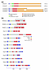
PHIST phylogeny and domain map. The PHIST tree (tree topology determined by bootstrapped neighbour joining based upon pairwise distances between instances of the PHIST domain; branch lengths assigned with least squares error minimisation; branches with <50% bootstrap support in red, 50% to 75% in blue, >75% in black) demonstrates conservation of the domain across Plasmodia. Colours indicate subfamilies (as determined by recognition by subfamily HMMs) and species conservation. Domain diagrams indicate organisational differences between subfamilies. The PHIST domain is carboxy-terminal in the PHISTc subfamily, regardless of length. In the PHISTa and b subfamilies a domain position of 100 to 200 amino acids from the amino-terminal methionine appears to be a general rule. In all the DnaJ containing members of the PHISTb subfamily, the DnaJ domain is carboxy-terminal to the PHIST domain. Members of the PHISTa subfamiliy are the shortest members of the PHIST family and are, as a whole, the most divergent and appear in a number of instances to be truncated. Note that PHIST domain representation is based on the annotated PlasmoDB [34] sequence, which in some cases lacks the first exon (for example, PFB0905c).
Figure 6
Figure 6
DNAJ phylogeny and gene/domain models. (a) The inset shows the complete tree of DnaJ domain sequences extracted from different Plasmodium species and C. hominis. Subtrees containing domains from exported (b) type I and (c) type III proteins are highlighted. DnaJ subtrees (type I, bootstrapped neighbour joining on full sequences; type III, TREE-PUZZLE on full sequences; branches with <50% bootstrap support in red, 50% to 75% in blue, >75% in black) show export (blue boxes) and core motif variations. Sequences not belonging to P. falciparum are marked with symbols to indicate the species group from which they arise. PF14_0359 has orthologs in other Plasmodia (not shown). Positions of signal sequence and PEXEL as determined by ExportPred, and PHIST and DnaJ domains as determined by HMM search, are overlaid on gene models. Although the large number of family members tends to decrease bootstrap support for individual branches, congruence with the three PHIST subfamilies as defined by HMM profiles lends additional support to the overall topology of the tree.
Figure 7
Figure 7
Plasmodium KAHRP orthologs and structural similarities of exported proteins. (a) Structural alignment of P. falciparum KAHRP and orthologs from P. fragile and P. vivax. Note that PfrKAHRP and PvKAHRP-c represent incomplete sequences (amino terminus represented as dashed line). (b) Structural representation of P. falciparum exported protein families. Secondary structure prediction was performed using the consensus method Jnet [84,85].

References

    1. Waller KL, Cooke BM, Nunomura W, Mohandas N, Coppel RL. Mapping the binding domains involved in the interaction between the Plasmodium falciparum knob-associated histidine-rich protein (KAHRP) and the cytoadherence ligand P. falciparum erythrocyte membrane protein 1 (PfEMP1). J Biol Chem. 1999;274:23808–23813. doi: 10.1074/jbc.274.34.23808. - DOI - PubMed
    1. Freitas-Junior LH, Hernandez-Rivas R, Ralph SA, Montiel-Condado D, Ruvalcaba-Salazar OK, Rojas-Meza AP, Mancio-Silva L, Leal-Silvestre RJ, Gontijo AM, Shorte S, Scherf A. Telomeric heterochromatin propagation and histone acetylation control mutually exclusive expression of antigenic variation genes in malaria parasites. Cell. 2005;121:25–36. doi: 10.1016/j.cell.2005.01.037. - DOI - PubMed
    1. Duraisingh MT, Voss TS, Marty AJ, Duffy MF, Good RT, Thompson JK, Freitas-Junior LH, Scherf A, Crabb BS, Cowman AF. Heterochromatin silencing and locus repositioning linked to regulation of virulence genes in Plasmodium falciparum. Cell. 2005;121:13–24. doi: 10.1016/j.cell.2005.01.036. - DOI - PubMed
    1. Chen Q, Schlichtherle M, Wahlgren M. Molecular aspects of severe malaria. Clin Microbiol Rev. 2000;13:439–450. doi: 10.1128/CMR.13.3.439-450.2000. - DOI - PMC - PubMed
    1. Kyes SA, Rowe JA, Kriek N, Newbold CI. Rifins: a second family of clonally variant proteins expressed on the surface of red cells infected with Plasmodium falciparum. Proc Natl Acad Sci USA. 1999;96:9333–9338. doi: 10.1073/pnas.96.16.9333. - DOI - PMC - PubMed

Publication types

Substances