. 2006;8(2):R49.
doi: 10.1186/ar1911. Epub 2006 Feb 28.

Abnormal costimulatory phenotype and function of dendritic cells before and after the onset of severe murine lupus

Affiliations

Abnormal costimulatory phenotype and function of dendritic cells before and after the onset of severe murine lupus

Lucrezia Colonna et al. Arthritis Res Ther. 2006.

Abstract

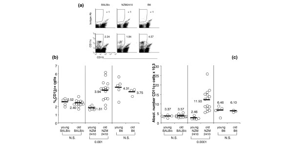
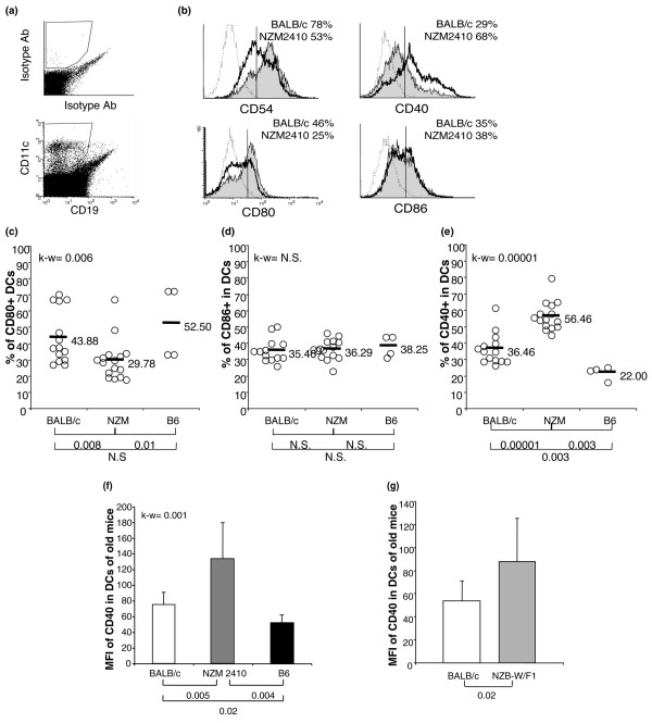
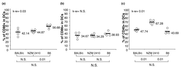
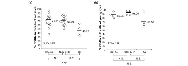
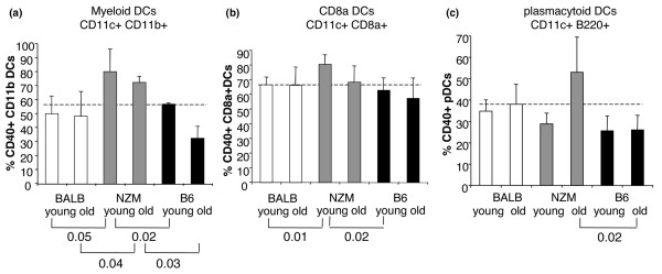
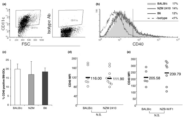
We analyzed the activation and function of dendritic cells (DCs) in the spleens of diseased, lupus-prone NZM2410 and NZB-W/F1 mice and age-matched BALB/c and C57BL/6 control mice. Lupus DCs showed an altered ex vivo costimulatory profile, with a significant increase in the expression of CD40, decreased expression of CD80 and CD54, and normal expression of CD86. DCs from young lupus-prone NZM2410 mice, before the development of the disease, expressed normal levels of CD80 and CD86 but already overexpressed CD40. The increase in CD40-positive cells was specific for DCs and involved the subset of myeloid and CD8alpha+ DCs before disease onset, with a small involvement of plasmacytoid DCs in diseased mice. In vitro data from bone marrow-derived DCs and splenic myeloid DCs suggest that the overexpression of CD40 is not due to a primary alteration of CD40 regulation in DCs but rather to an extrinsic stimulus. Our analyses suggest that the defect of CD80 in NZM2410 and NZB-W/F1 mice, which closely resembles the costimulatory defect found in DCs from humans with systemic lupus erythematosus, is linked to the autoimmune disease. The increase in CD40 may instead participate in disease pathogenesis, being present months before any sign of autoimmunity, and its downregulation should be explored as an alternative to treatment with anti-CD40 ligand in lupus.


Figures

Figure 1
Figure 1
DCs accumulate with age in mice with lupus. (a) To study DCs, we analyzed the total population of splenocytes in mice 6 to 8 weeks old (young) and in mice 6 to 9 months old (old) and gated for CD11c+ cells that were negative for the B cell marker CD19. (b,c) Percentages (b) and absolute numbers (c) of DCs gated as in (a). Each circle represents the percentage or absolute number of CD11c+ cells in one individual mouse, and the horizontal bars represent the average of each group. Results of the Mann-Whitney test analyses are shown. N.S. indicates p > 0.05.
Figure 2
Figure 2
DCs express an altered costimulatory phenotype in lupus diseased mice. (a) To study DCs, we analyzed the total population of splenocytes in mice 6 to 9 months old and gated for CD11c+ cells that were negative for the B-cell marker CD19. (b) DCs from mice with lupus (thick dark histograms) express lower levels of CD80, normal levels of CD86, and higher levels of CD40 than DCs of non-autoimmune mice (grey filled histograms). The highlighted numbers are the percentages of DCs positive for the indicated markers and are representative of several mice analyzed and shown in (c-e). The vertical line delineates the threshold of positivity, set on the isotype control background less than 1% (dotted histograms). (c-e) Positivity for CD80 (c), CD86 (d), and CD40 (e) in DCs gated as in (a). Each circle represents the percentage of positive cells in one individual mouse, and the horizontal bars represent the average of each group. (f,g) Averages and standard deviations of CD40 mean fluorescence intensity for expression in DCs from mice 6 to 9 months old of the indicated strains. We analyzed four to nine mice per group in (f) and seven to nine mice per group in (g). Results of the Kruskal-Wallis test analyses are shown in the graphs (k-w) and the post hoc Mann-Whitney test analyses are shown below the graphs. N.S. indicates p > 0.05.
Figure 3
Figure 3
Increase in CD40-positive DCs pre-dates lupus onset of disease, whereas CD80/CD86 balance is normal. Percentages of cells positive for CD80 (a), CD86 (b), and CD40 (c) in DCs from spleens of young (6 to 8 weeks old) mice. We analyzed DCs as described in Figure 2. Results of the Kruskal-Wallis test analyses are shown in the graphs (k-w) and the post hoc Mann-Whitney test analyses are shown below the graphs. N.S. indicates p > 0.05.
Figure 4
Figure 4
B cells from lupus-prone mice express normal levels of CD40. Percentages of B cells positive for CD40 from mice 6 to 9 months old (a) and 6 to 8 weeks old (b) are shown. B cells were gated as B220/CD19-positive and CD11c-negative cells. Results of the Kruskal-Wallis test analyses are shown in the graphs (k-w) and the post hoc Mann-Whitney test analyses are shown below the graphs. N.S. indicates p > 0.05.
Figure 5
Figure 5
CD40 expression in three major DC subsets. Percentages and standard deviations of CD40-positive cells in myeloid (a), CD8α+ (b), and plasmacytoid (c) DC subsets from young (6 to 8 weeks old) and old (6 to 9 months old) mice were calculated from four to nine mice per group. Only results by the Mann-Whitney test analyses with p < 0.05 are indicated.
Figure 6
Figure 6
Regulation of CD40 expression is not intrinsically altered in lupus DCs. We grew bone marrow-derived dendritic cells (BM-DCs) from NZM2410, BALB/c, and B6 mice 6 to 9 months old and stained them at day 6 to 7 of culture. Gating of CD11c+ cells (a) and CD40 expression (b) in resting BM-DCs from NZM2410 (thick dark grey line), BALB/c (light grey area), and B6 (thin black line) mice. (c) Percentages of CD40-positive BM-DCs, gated as in (a) and considered CD40-positive using the threshold shown in (b) from three experiments, conducted with three independent BM-DCs cultures. Error bars show SD. (d,e) Mean fluorescence intensity (MFI) of CD40 expression in splenic myeloid DCs isolated and cultured overnight in vitro from NZM2410 (d) and NZB-W/F1 (e) mice 6 to 9 months old.

References

    1. Cederblad B, Blomberg S, Vallin H, Perers A, Alm GV, Ronnblom L. Patients with systemic lupus erythematosus have reduced numbers of circulating natural interferon-alpha-producing cells. J Autoimmun. 1998;11:465–470. doi: 10.1006/jaut.1998.0215. - DOI - PubMed
    1. Garcia-Cozar FJ, Molina IJ, Cuadrado MJ, Marubayashi M, Pena J, Santamaria M. Defective B7 expression on antigen-presenting cells underlying T cell activation abnormalities in systemic lupus erythematosus (SLE) patients. Clin Exp Immunol. 1996;104:72–79. doi: 10.1046/j.1365-2249.1996.d01-648.x. - DOI - PMC - PubMed
    1. Liu MF, Li JS, Weng TH, Lei HY. Differential expression and modulation of costimulatory molecules CD80 and CD86 on monocytes from patients with systemic lupus erythematosus. Scand J Immunol. 1999;49:82–87. doi: 10.1046/j.1365-3083.1999.00452.x. - DOI - PubMed
    1. Funauchi M, Yoo BS, Nozaki Y, Sugiyama M, Ohno M, Kinoshita K, Kanamaru A. Dysregulation of the granulocyte-macrophage colony-stimulating factor receptor is one of the causes of defective expression of CD80 antigen in systemic lupus erythematosus. Lupus. 2002;11:317–321. doi: 10.1191/0961203302lu201oa. - DOI - PubMed
    1. Blanco P, Palucka AK, Gill M, Pascual V, Banchereau J. Induction of dendritic cell differentiation by IFN-α in systemic lupus erythematosus. Science. 2001;294:1540–1543. doi: 10.1126/science.1064890. - DOI - PubMed

Publication types