. 2006;8(2):R50.
doi: 10.1186/ar1913. Epub 2006 Feb 28.

Expression of the inflammatory chemokines CCL5, CCL3 and CXCL10 in juvenile idiopathic arthritis, and demonstration of CCL5 production by an atypical subset of CD8+ T cells

Affiliations

Expression of the inflammatory chemokines CCL5, CCL3 and CXCL10 in juvenile idiopathic arthritis, and demonstration of CCL5 production by an atypical subset of CD8+ T cells

Daniel S Pharoah et al. Arthritis Res Ther. 2006.

Abstract

This study focuses upon three chemokines, namely CCL5, CXCL10 and CCL3, which are potential novel therapeutic targets in arthritis. The aim of the study was to analyse the expression and production of these three chemokines within the joints of children with juvenile idiopathic arthritis (JIA) of the oligoarticular and polyarticular subtypes. All three of these chemokines are highly expressed at the level of mRNA, with the most significant increase in mRNA levels being demonstrated for CCL5 when compared with matched peripheral blood samples and controls. We show that high levels of all three chemokines are present in synovial fluid of children with JIA. We investigate the major source of CCL5 from inflammatory synovial cells, which we show to be CD8+ T cells. This CD8+ synovial T cell population has an unexpected phenotype that has not been described previously, being CCR7- yet predominantly CD28+ and CD45RA-. These cells contain high levels of stored intracellular CCL5, and rapid release of CCL5 takes place on T cell stimulation, without requiring new protein synthesis. In addition, we demonstrate that CCL5 is present in synovial biopsies from these patients, in particular on the endothelium of small and medium sized vessels. We believe this to be the first in depth analysis of these mediators of inflammation in JIA.


Figures

Figure 1
Figure 1
Expression of mRNA for three inflammatory chemokines in juvenile idiopathic arthritis. (a) Amplification products of mRNA for CCL5, CCL3 and CXCL10 after RT-PCR from peripheral blood (PB) and synovial fluid (SF) mononuclear cells from patients (n = 8) with juvenile idiopathic arthritis (JIA; 5 persistent oligoarticular, 3 extended oligoarticular) and 5 controls. Amplification of β-actin acted as control. (b) Amplification of mRNA for CCL3, CXCL10 and β-actin from myeloid (M) or lymphoid (L) cells purified from three representative JIA patients (one persistent oligoarticular, one extended oligoarticular, and one polyarticular).
Figure 2
Figure 2
High expression of mRNA for CCL5 in purified T cells from the joint of patients with juvenile idiopathic arthritis (JIA). (a) Oligonucleotide-specific probing of blotted amplification products of CCL5 after RT-PCR from paired sets of purified T cells from 15 JIA patients (5 persistent oligoarticular, 5 extended oligoarticular, 5 polyarticular). (b) Relative levels of CCL5 mRNA in purified T cells from each JIA subgroup. Black bars, peripheral blood (PB) CD3+ cells; white bars, synovial fluid (SF) CD3+ cells. Bars represent mean values; error bars one standard deviation.
Figure 3
Figure 3
Intracellular flow cytometric analysis of CCL5+ synovial T cells. Peripheral blood (PB) and synovial cells were stained for CD3, CD8 and CCL5. (a) Histograms represent CCL5 expression in cells from PB (thin line), or synovial fluid (SF; bold line), compared to staining by isotype control (dashed line), with events gated either on all CD3+ cells (left panel), or CD3+CD8+ cells only (right panel). Marker (M1) indicates CCL5 positive cells. (b) Summary of flow cytometric analysis of CCL5 expression from 11 JIA patients (4 persistent oligoarticular, closed triangles; 5 extended oligoarticular, closed circles; and 2 polyarticular, closed squares), indicating number of CD3+CD8+ cells staining positive for CCL5 in PB and SF.
Figure 4
Figure 4
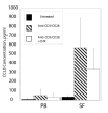
Release of CCL5 from synovial cells is very rapid and largely independent of new protein synthesis. Purified T cells from peripheral blood (PB) and synovial fluid (SF) were stimulated with anti-CD3 and anti-CD28, with (clear bars) or without (hatched bars) cyclohexamide (to block protein synthesis), or in control medium (black bars). Supernatants were assayed by standard ELISA for CCL5. Bars represent mean values of CCL5, and error bars one standard deviation.
Figure 5
Figure 5
The phenotype of CCL5+ synovial T cells diverges from the effector memory phenotype. Flow cytometric analysis of peripheral blood (PB) and synovial cells. (a) Cells were stained for CD3, CD8 and CR7 and CD28. The majority of synovial fluid (SF) T cells were CCR7- (upper panels). However, within the CCR7- population of CD8+T cells, synovial T cells showed maintained high expression of CD28 compared to CD8+CCR7- T cells from PB (lower panel). (b) Costaining for CD8, CCR7, CCL5 and CD28 confirmed that the synovial CCR7-CD28+CD8+ cells also contained high levels of intracellular CCL5; positive staining for CCL5 was defined by comparison with isotype-matched control staining. (c) The pattern of CCL5+ staining seen in CD8+CD28+ T cells was seen in a total of 11 sets of samples (4 persistent oligoarticular, closed triangles; 5 extended oligoarticular, closed circles; and 2 polyarticular, closed squares), when comparing cells from PB and SF.
Figure 6
Figure 6
Intense staining for CCL5 on endothelium within inflamed synovial tissue of juvenile idiopathic arthritis patients. Immuno-staining for (a) CCL5 on synovial biopsy tissue taken from a child with oligoarticular juvenile idiopathic arthritis, carried out on a frozen section by standard immunohistochemistry for (b) CCL5 or with isotype control (both images 100×). CCL5 is seen to be expressed on endothelial cells and infiltrating inflammatory cells.

References

    1. Bywaters EG. Pathologic aspects of juvenile chronic polyarthritis. Arthritis Rheum. 1977;20:271–276. - PubMed
    1. Wynne-Roberts CR, Anderson CH, Turano AM, Baron M. Light- and electron-microscopic findings of juvenile rheumatoid arthritis synovium: comparison with normal juvenile synovium. Semin Arthritis Rheum. 1978;7:287–302. doi: 10.1016/0049-0172(78)90027-6. - DOI - PubMed
    1. Wedderburn LR, Maini MK, Patel A, Beverley PCL, Woo P. Molecular fingerprinting reveals non-overlapping T cell oligoclonality between an inflamed site and peripheral blood. Int Immunol. 1999;11:535–543. doi: 10.1093/intimm/11.4.535. - DOI - PubMed
    1. Butcher EC, Scollay RG, Weissman IL. Organ specificity of lymphocyte migration: mediation by highly selective lymphocyte interaction with organ-specific determinants on high endothelial venules. Eur J Immunol. 1980;10:556–561. - PubMed
    1. Jalkanen S, Reichert RA, Gallatin WM, Bargatze RF, Weissman IL, Butcher EC. Homing receptors and the control of lymphocyte migration. Immunol Rev. 1986;91:39–60. doi: 10.1111/j.1600-065X.1986.tb01483.x. - DOI - PubMed

Publication types

MeSH terms