. 2006 Jun 5;349(2):264-75.
doi: 10.1016/j.virol.2006.01.028. Epub 2006 Feb 28.

Biochemical and functional characterization of the membrane association and membrane permeabilizing activity of the severe acute respiratory syndrome coronavirus envelope protein

Affiliations

Biochemical and functional characterization of the membrane association and membrane permeabilizing activity of the severe acute respiratory syndrome coronavirus envelope protein

Y Liao et al. Virology. .

Abstract

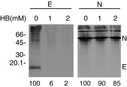
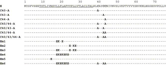
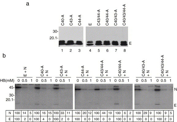
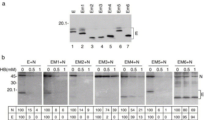
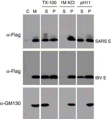
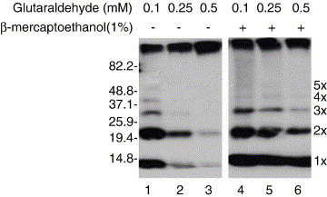
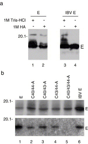
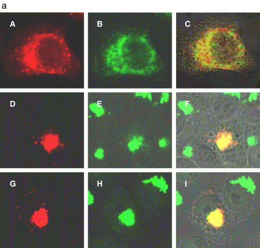
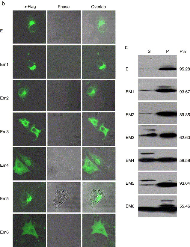
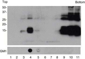
A diverse group of cytolytic animal viruses encodes small, hydrophobic proteins to modify host cell membrane permeability to ions and small molecules during their infection cycles. In this study, we show that expression of the SARS-CoV E protein in mammalian cells alters the membrane permeability of these cells. Immunofluorescent staining and cell fractionation studies demonstrate that this protein is an integral membrane protein. It is mainly localized to the ER and the Golgi apparatus. The protein can be translocated to the cell surface and is partially associated with lipid rafts. Further biochemical characterization of the protein reveals that it is posttranslationally modified by palmitoylation on all three cysteine residues. Systematic mutagenesis studies confirm that the membrane permeabilizing activity of the SARS-CoV E protein is associated with its transmembrane domain.


Figures

Fig. 1
Fig. 1
Modification of the membrane permeability of mammalian cells by SARS-CoV E protein. HeLa cells expressing the Flag-tagged E protein were treated with 0, 1, and 2 mM of hygromycin B for 30 min at 12 h posttransfection (lanes 1, 2, and 3), and radiolabeled with [35S] methionine–cysteine for 3 h. Cell lysates were prepared and the expression of E protein was detected by immunoprecipitation with anti-Flag antibody under mild washing conditions. Polypeptides were separated by SDS-PAGE and visualized by autoradiography. Cells expressing SARS-CoV N proteins were included as negative control (lanes 4, 5, and 6). The expression of N protein was detected by immunoprecipitation with polyclonal anti-N antibodies. The percentages of E and N proteins detected in the presence of hygromycin B were determined by densitometry and indicated at the bottom. Numbers on the left indicate molecular masses in kilodaltons.
Fig. 2
Fig. 2
Amino acid sequences of wild type and mutant SARS-CoV E protein. The putative transmembrane domain is underlined, and the three cysteine residues are in bold. Also indicated are the amino acid substitutions in each mutant construct.
Fig. 3
Fig. 3
Mutational analysis of the three cysteine residues of SARS-CoV E protein. (a) HeLa cells were transfected with the Flag-tagged wild type and seven mutant E constructs containing mutations of either a single (C40-A, C43-A, and C44-A), combination of two (C40/43-A, C40/44-A, and C43/44-A) or all three (C40/43/44-A) cysteine residues. Cell lysates were prepared at 24 h posttransfection, polypeptides were separated by SDS-PAGE and analyzed by Western blot using the anti-Flag antibody. Numbers on the left indicate molecular masses in kilodaltons. (b) Entry of hygromycin B into HeLa cells expressing wild type and mutant E proteins. HeLa cells expressing the Flag-tagged wild type E (lanes 1, 2, and 3) and seven cysteine to alanine mutation constructs (lanes 4–24) were treated with 0, 0.5, and 1 mM of hygromycin B for 30 min at 12 h posttransfection, and radiolabeled with [35S] methionine–cysteine for 3 h. Cell lysates were prepared and the expression of E protein was detected by immunoprecipitation with anti-Flag antibody under mild washing conditions. SARS-CoV N protein was coexpressed with wild type and mutant E protein, and the expression of N protein was detected by immunoprecipitation with polyclonal anti-N antibodies. Polypeptides were separated by SDS-PAGE and visualized by autoradiography. The percentages of E and N proteins detected in the presence of hygromycin B were determined by densitometry and indicated at the bottom. Numbers on the left indicate molecular masses in kilodaltons.
Fig. 4
Fig. 4
Mutational analysis of the transmembrane domain of SARS-CoV E protein. (a) HeLa cells were transfected with the Flag-tagged wild type and six mutant constructs containing mutations in the transmembrane domain of the E protein. Cell lysates were prepared 24 h posttransfection, polypeptides were separated by SDS-PAGE and analyzed by Western blot using the anti-Flag antibody. Numbers on the left indicate molecular masses in kilodaltons. (b) Entry of hygromycin B into HeLa cells expressing wild type and mutant E proteins. HeLa cells expressing the Flag-tagged wild type E (lanes 1, 2, and 3) and six mutant E constructs (lanes 4–21), respectively, were treated with 0, 0.5, and 1 mM of hygromycin B for 30 min at 12 h posttransfection, and radiolabeled with [35S] methionine–cysteine for 3 h. Cell lysates were prepared and the expression of E protein was detected by immunoprecipitation with anti-Flag antibody under mild washing conditions. SARS-CoV N protein was coexpressed with wild type and mutant E protein, and the expression of N protein was detected by immunoprecipitation with polyclonal anti-N antibodies. Polypeptides were separated by SDS-PAGE and visualized by autoradiography. The percentages of E and N proteins detected in the presence of hygromycin B were determined by densitometry and indicated at the bottom. Numbers on the left indicate molecular masses in kilodaltons.
Fig. 5
Fig. 5
Determination of SARS-CoV E protein as an integral membrane protein. HeLa cells expressing the Flag-tagged SARS-CoV and IBV E proteins, respectively, were harvested at 12 h posttransfection, broken by 20 stokes with a Dounce cell homogenizer, and fractionated into cytosol (C) and membrane (M) fractions after removal of cell debris and nuclei. The membrane fraction was treated with 1% Triton X-100, 100 mM Na2CO3 (pH 11), and 1 M KCl, respectively, and further fractionated into soluble (S) and pellet (P) fractions. Polypeptides were separated by SDS-PAGE and analyzed by Western blot using either anti-Flag antibody or anti-GM130 antibody (Abcam). Numbers on the left indicate molecular masses in kilodaltons.
Fig. 6
Fig. 6
Oligomerization of SARS-CoV E protein. The His-tagged E protein expressed in Sf9 insect cells was purified using Ni-NTA purification system (Qiagen), and incubated with three different concentrations of glutaraldehyde (0.1, 0.25, and 0.5 mM) for 1 h at room temperature. The reaction was quenched by adding 100 mM glycine. Polypeptides were separated on SDS-15% polyacrylamide gel in the presence or absence of 1% β-mercaptoethanol, and analyzed by Western blot with anti-His antibody. Different oligomers of the E protein are indicated on the right. Numbers on the left indicate molecular masses in kilodaltons.
Fig. 7
Fig. 7
Palmitoylation of SARS-CoV E protein. (a) Total cell lysates prepared from HeLa cells expressing the Flag-tagged SARS-CoV E protein (lanes 1 and 2) and IBV E protein (lanes 3 and 4) were treated either with 1 M Tris–HCl (lanes 1 and 3) or 1M hydroxylamine (lanes 2 and 4). Polypeptides were separated by SDS-PAGE and analyzed by Western blot using the anti-Flag antibody. Numbers on the left indicate molecular masses in kilodaltons. (b) HeLa cells expressing the Flag-tagged wild type SARS-CoV E protein (lane 1), mutant C40/44-A (lane 2), C40/44-A (lane 3), C43/44-A (lane4), C40/43/44-A (lane 5), and IBV E protein (lane 6) were radiolabeled with [35S] methionine–cysteine (upper panel) and [3H] palmitic acid (lower panel). Cell lysates were prepared and subjected to immunoprecipitation with anti-Flag antibody. Polypeptides were separated by SDS-PAGE and visualized by autoradiography. Numbers on the left indicate molecular masses in kilodaltons.
Fig. 8
Fig. 8
Subcellular localization and membrane association of wild type and mutant SARS-CoV E protein. (a) HeLa cells expressing the Flag-tagged E protein (A–C), and BHK cells expressing the Flag-tagged (D–F) and untagged (G–I) SARS-CoV E were stained with either anti-Flag (A–F) or anti-E (G–I) antibodies at 12 h posttransfection after permeabilizing with 0.2% Triton X-100. The same HeLa cells were also stained with anti-calnexin antibody (B), and the same BHK cells were also stained with anti-p230 trans Golgi antibodies (panels E and H). Panels C, F, and I show the overlapping images. (b) BHK cells expressing the Flag-tagged wild type (E) and mutant E protein (Em1, Em2, Em3, Em4, Em5, and Em6) were stained with anti-Flag antibody at 12 h posttransfection after permeabilizing with 0.2% Triton X-100. (c) HeLa cells expressing the Flag-tagged wild type and mutant E protein were harvested at 12 h posttransfection, broken by 20 stokes with a Dounce cell homogenizer, and fractionated into cytosol (C) and membrane (M) fractions after removal of cell debris and nuclei. Polypeptides were separated by SDS-PAGE and analyzed by Western blot using the anti-Flag antibody. The percentages of E protein detected in the membrane fraction were determined by densitometry and indicated on the right. Numbers on the left indicate molecular masses in kilodaltons.
Fig. 8
Fig. 8
Subcellular localization and membrane association of wild type and mutant SARS-CoV E protein. (a) HeLa cells expressing the Flag-tagged E protein (A–C), and BHK cells expressing the Flag-tagged (D–F) and untagged (G–I) SARS-CoV E were stained with either anti-Flag (A–F) or anti-E (G–I) antibodies at 12 h posttransfection after permeabilizing with 0.2% Triton X-100. The same HeLa cells were also stained with anti-calnexin antibody (B), and the same BHK cells were also stained with anti-p230 trans Golgi antibodies (panels E and H). Panels C, F, and I show the overlapping images. (b) BHK cells expressing the Flag-tagged wild type (E) and mutant E protein (Em1, Em2, Em3, Em4, Em5, and Em6) were stained with anti-Flag antibody at 12 h posttransfection after permeabilizing with 0.2% Triton X-100. (c) HeLa cells expressing the Flag-tagged wild type and mutant E protein were harvested at 12 h posttransfection, broken by 20 stokes with a Dounce cell homogenizer, and fractionated into cytosol (C) and membrane (M) fractions after removal of cell debris and nuclei. Polypeptides were separated by SDS-PAGE and analyzed by Western blot using the anti-Flag antibody. The percentages of E protein detected in the membrane fraction were determined by densitometry and indicated on the right. Numbers on the left indicate molecular masses in kilodaltons.
Fig. 9
Fig. 9
Association of SARS-CoV E protein with lipid rafts. HeLa cells expressing the Flag-tagged SARS E protein were lysed with 1% Triton, and centrifuged to remove insoluble material and nuclei. The supernatants were fractionated by ultracentrifugation with a sucrose gradient, and 11 fractions were collected. The presence of the SARS-CoV E protein in each fraction was analyzed by Western blot using anti-Flag antibody, and the presence of GM1 was determined by dot blot. Numbers on the left indicate molecular masses in kilodaltons.

Similar articles

Cited by

References

    1. Agirre A., Barco A., Carrasco L., Nieva J.B. Viroporin-mediated membrane permeabilization. J. Biol. Chem. 2002;277:40434–40441. - PubMed
    1. Aldabe R., Barco A., Corrasco L. Membrane permeabilization by poliovirus proteins 2B and 2BC. J. Biol. Chem. 1996;271:23134–23137. - PubMed
    1. Arbely E., Khattari Z., Brotons G., Akkawi M., Salditt T., Arkin I.T. A highly unusual palindromic transmembrane helical hairpin formed by SARS coronavirus E protein. J. Mol. Biol. 2004;341:769–779. - PMC - PubMed
    1. Bhattacharya J., Peters P.J., Clapham P.R. Human immunodeficiency virus type 1 envelope glycoproteins that lack cytoplasmic domain cysteines: impact on association with membrane lipid rafts and incorporation onto budding virus particles. J. Virol. 2004;78:5500–5506. - PMC - PubMed
    1. Bijlmakers M.-J., Marsh M. The on–off story of protein palmitoylation. Trends Cell Biol. 2003;13:32–42. - PubMed

Publication types

MeSH terms