. 2006 Mar;114(3):412-9.
doi: 10.1289/ehp.8413.

Systemic microvascular dysfunction and inflammation after pulmonary particulate matter exposure

Affiliations

Systemic microvascular dysfunction and inflammation after pulmonary particulate matter exposure

Timothy R Nurkiewicz et al. Environ Health Perspect. 2006 Mar.

Abstract

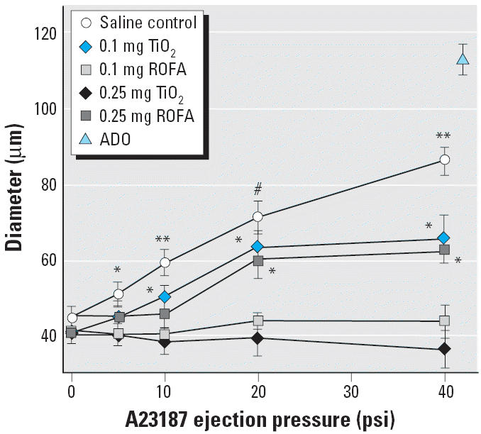
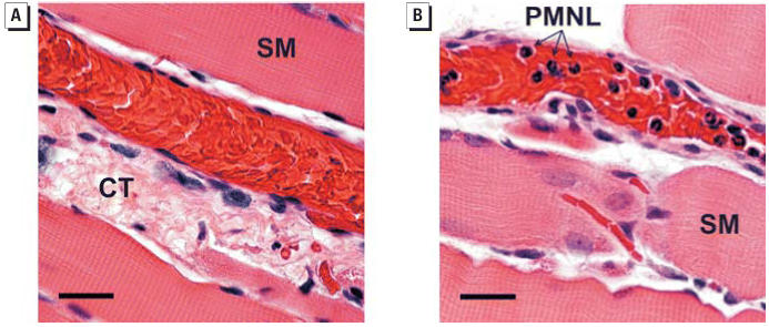
The epidemiologic association between pulmonary exposure to ambient particulate matter (PM) and cardiovascular dysfunction is well known, but the systemic mechanisms that drive this effect remain unclear. We have previously shown that acute pulmonary exposure to PM impairs or abolishes endothelium-dependent arteriolar dilation in the rat spinotrapezius muscle. The purpose of this study was to further characterize the effect of pulmonary PM exposure on systemic microvascular function and to identify local inflammatory events that may contribute to these effects. Rats were intratracheally instilled with residual oil fly ash (ROFA) or titanium dioxide at 0.1 or 0.25 mg/rat 24 hr before measurement of pulmonary and systemic microvascular responses. In vivo microscopy of the spinotrapezius muscle was used to study systemic arteriolar responses to intraluminal infusion of the Ca2+ ionophore A23187 or iontophoretic abluminal application of the adrenergic agonist phenylephrine (PHE). Leukocyte rolling and adhesion were quantified in venules paired with the studied arterioles. Histologic techniques were used to assess pulmonary inflammation, characterize the adherence of leukocytes to systemic venules, verify the presence of myeloperoxidase (MPO) in the systemic microvascular wall, and quantify systemic microvascular oxidative stress. In the lungs of rats exposed to ROFA or TiO2, changes in some bronchoalveolar lavage markers of inflammation were noted, but an indication of cellular damage was not found. In rats exposed to 0.1 mg ROFA, focal alveolitis was evident, particularly at sites of particle deposition. Exposure to either ROFA or TiO2 caused a dose-dependent impairment of endothelium-dependent arteriolar dilation. However, exposure to these particles did not affect microvascular constriction in response to PHE. ROFA and TiO2 exposure significantly increased leukocyte rolling and adhesion in paired venules, and these cells were positively identified as polymorphonuclear leukocytes (PMNLs). In ROFA- and TiO2-exposed rats, MPO was found in PMNLs adhering to the systemic microvascular wall. Evidence suggests that some of this MPO had been deposited in the microvascular wall. There was also evidence for oxidative stress in the microvascular wall. These results indicate that after PM exposure, the impairment of endothelium-dependent dilation in the systemic microcirculation coincides with PMNL adhesion, MPO deposition, and local oxidative stress. Collectively, these microvascular observations are consistent with events that contribute to the disruption of the control of peripheral resistance and/or cardiac dysfunction associated with PM exposure.


Figures

Figure 1
Figure 1
Histologic evidence of focal pulmonary alveolitis 24 hr after PM exposure. (A) and (B) are representative findings from five saline-treated rats and five rats exposed to 0.1 mg ROFA. (A) Saline control showing no morphologic alterations. Abbreviations: AD, alveolar duct; TB, terminal bronchioles. Bar = 50 μm. (B) Histopathologic alterations in a ROFA-exposed rat. Agglomerated ROFA particles in an alveolar space can be seen near an alveolar duct. The ROFA particles do not transmit light and therefore appear black when viewed in the light microscope. AMs are frequently observed in alveoli near ROFA particles and are intimately associated with the agglomerated ROFA. PMNLs are present in lesser numbers near ROFA particles, most frequently in the interstitium. Bar = 20 μm. (C) Mean alveolitis pathology scores. *p < 0.05 compared with saline; similar results were obtained with TiO2.
Figure 2
Figure 2
PM exposure impairs or abolishes spinotrapezius muscle arteriolar responsiveness to intraluminal A23187 in a dose-dependent manner 24 hr after IT treatment. n, number of arterioles. Saline control, n = 13; 0.1 mg TiO2, n = 9; 0.1 mg ROFA, n = 9; 0.25 mg TiO2, n = 8; 0.25 mg ROFA, n = 8. Maximum diameter obtained with ADO (10−4 M, final superfusate concentration). Values are mean ± SE. *p < 0.05 compared with 0.25 mg TiO2 and 0.25 mg ROFA. **p < 0.05 compared with 0.1 mg TiO2, 0.1 mg ROFA, 0.25 mg TiO2, and 0.25 mg ROFA. #p < 0.05 compared with 0.1 mg ROFA, 0.25 mg TiO2, and 0.25 mg ROFA.
Figure 3
Figure 3
ROFA exposure does not alter arteriolar vasopressor responsiveness to PHE in the spinotrapezius muscle. n, number of arterioles. Saline control, n = 8; 0.25 mg ROFA, n = 8. Values are mean ± SE. *p < 0.05 compared with 0 nA in both groups; **p < 0.05 compared with 50 nA in both groups; #p < 0.05 compared with 100 nA in both groups.
Figure 4
Figure 4
PM exposure increases venular leukocyte rolling and adhesion in the spinotrapezius muscle 24 hr after IT treatment. Venular leukocytes were quantified as rolling and adhering leukocytes per minute in a 200-μm segment. n, number of venules. Saline control, n = 26; 2 mg ROFA, n = 15; 0.25 mg ROFA, n = 18; 0.1 mg TiO2, n = 10. Values are mean ± SE. *p < 0.05 compared with saline control.
Figure 5
Figure 5
PMNL identification in the spinotrapezius muscle microcirculation of PM-exposed rats 24 hr after IT exposure. (A) Representative H&E-stained section from a saline-treated rat. Abbreviations: CT, connective tissue; SM, skeletal muscle fiber. (B) Representative H&E-stained section from a rat exposed to 0.1 mg ROFA. Note the deeply lobed nuclei that are characteristic of PMNLs. Bars = 25 μm; similar results were obtained with TiO2.
Figure 6
Figure 6
Microvascular inflammatory markers 24 hr after exposure to ROFA: mRNA measurements in microvessels from six saline-treated rats and six rats exposed to 0.25 mg ROFA. After microdissection, vessels from both spinotrapezius muscles of an individual rat were pooled for data collection. Abbreviations: MIP-2, macrophage inflammatory protein; VCAM-1, vascular cell adhesion molecule. Note that neither eNOS nor iNOS message was altered after ROFA exposure.
Figure 7
Figure 7
Localization of MPO in the spinotrapezius muscle microcirculation 24 hr after ROFA exposure. Fluorescent antibodies targeted a polyclonal antibody against MPO; nuclei are counterstained blue with DAPI. (A) Representative confocal fluorescent image of a venule from a saline-treated rat. (B) Representative confocal image of a venule from a rat exposed to 0.25 mg ROFA. Note the fluorescence in the microvascular wall indicating the presence of MPO (arrows). Bars = 20 μm; similar results were obtained with TiO2.
Figure 8
Figure 8
ROFA exposure increases oxidative stress in the systemic microcirculation 24 hr after IT treatment. n = number of vessels. Calculated wall light absorption [A = ln(lt/lo)] in microvessels from saline-treated rats (n = 16) and rats exposed to 0.25 mg ROFA (n = 15) after exposure to 2% TNBT. Values are mean ± SE. *p < 0.05 compared with saline control.

Similar articles

Cited by

References

    1. Antonini JM, Roberts JR, Jernigan MR, Yang HM, Ma JY, Clarke RW. Residual oil fly ash increases the susceptibility to infection and severely damages the lungs after pulmonary challenge with a bacterial pathogen. Toxicol Sci. 2002;70:110–119. - PubMed
    1. Bagate K, Meiring JJ, Cassee FR, Borm PJ. The effect of particulate matter on resistance and conductance vessels in the rat. Inhal Toxicol. 2004;16:431–436. - PubMed
    1. Baldus S, Eiserich JP, Mani A, Castro L, Figueroa M, Chumley P, et al. Endothelial transcytosis of myeloperoxidase confers specificity to vascular ECM proteins as targets of tyrosine nitration. J Clin Invest. 2001;108:1759–1770. - PMC - PubMed
    1. Brain JD, Knudson DE, Sorokin SP, Davis MA. Pulmonary distribution of particles given by intratracheal instillation or by aerosol inhalation. Environ Res. 1976;11:13–33. - PubMed
    1. Brook RD, Brook JR, Urch B, Vincent R, Rajagopalan S, Silverman F. Inhalation of fine particulate air pollution and ozone causes acute arterial vasoconstriction in healthy adults. Circulation. 2002;105:1534–1536. - PubMed

Publication types

MeSH terms