. 2006 Mar;168(3):812-21.
doi: 10.2353/ajpath.2006.050923.

Inhibitory role of CD19 in the progression of experimental autoimmune encephalomyelitis by regulating cytokine response

Affiliations

Inhibitory role of CD19 in the progression of experimental autoimmune encephalomyelitis by regulating cytokine response

Takashi Matsushita et al. Am J Pathol. 2006 Mar.

Abstract

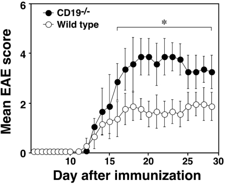
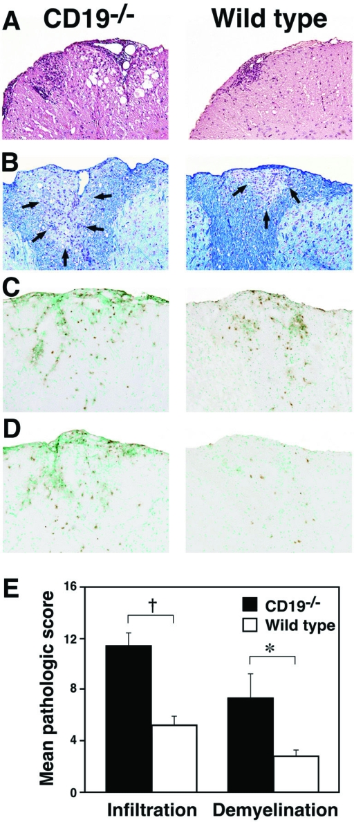
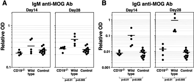
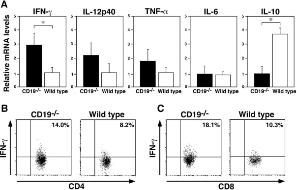
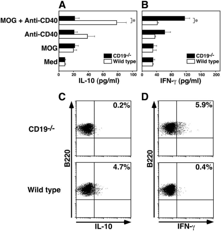
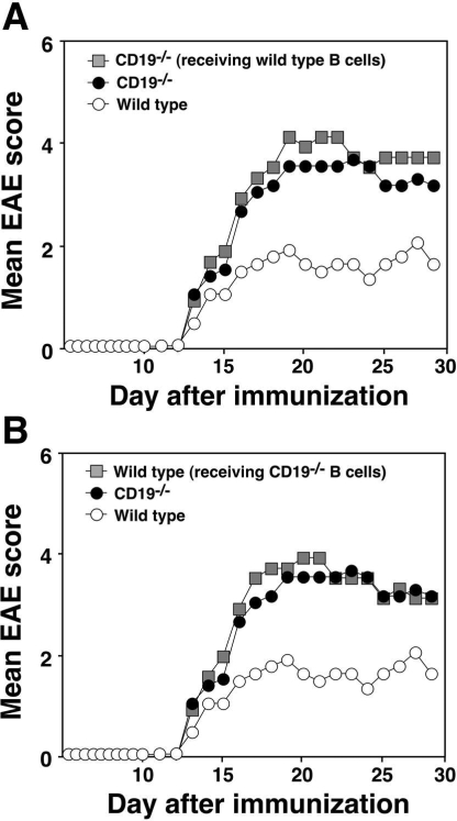
Experimental autoimmune encephalomyelitis (EAE) is an inflammatory demyelinating disease of the central nerve system that is considered a T helper type 1 (Th1)-mediated autoimmune disease. EAE currently serves as an experimental animal model for multiple sclerosis in human. Cytokines, such as interferon-gamma and interleukin-10, play a key role in the development and remission of EAE. Recent studies have also shown a role for B cells in the pathogenesis of EAE. Therefore, we examined the role of CD19, a B cell-specific surface molecule that defines signaling thresholds critical for B-cell responses and autoimmunity, on the development of EAE. Following immunization with myelin oligodendrocyte glycoprotein (MOG) peptide, CD19-deficient (CD19(-/-)) mice exhibited higher clinical and pathological severity scores of EAE than wild-type mice. The increased severity of EAE in CD19(-/-) mice was associated with polarized Th1 cytokines in the inflamed central nerve system but not with anti-MOG antibodies in the serum. MOG-primed CD19(-/-) B cells produced high levels of interferon-gamma, and transfer of MOG-primed CD19(-/-) B cells to wild-type mice worsened the disease. Thus, CD19 modulates the Th1/Th2 cytokine balance in B cells and plays a critical role as a suppressive molecule in the development of EAE.


Figures

Figure 1
Figure 1
Increased severity of EAE in CD19−/− mice. CD19−/− and wild-type mice (10 per group) were immunized with MOG peptide in CFA and intravenously injected with pertussis toxin. Mice were scored for the severity of EAE using the scale described in Materials and Methods. The EAE score is shown as mean ± SEM. *P < 0.05.
Figure 2
Figure 2
CD19−/− mice had higher pathological severity scores of EAE. Spinal cords were harvested from CD19−/− and wild-type mice (five per group) 28 days after immunization. A representative section from each group shows lymphocyte infiltration stained by H&E (A), demyelination stained by Luxol Fast Blue (B), immunohistochemistry for CD4+ T cells (C) and CD8+ T cells (D), and the mean pathological score of demyelination and infiltration of each group (E). Original magnifications: ×200. Arrows point to the foci of demyelination. Data are the mean ± SEM. *P < 0.05; P < 0.005.
Figure 3
Figure 3
CD19 loss attenuates anti-MOG Ig production. CD19−/− and wild-type mice were immunized with MOG peptide in CFA. Sera (five per group) were collected on days 14 and 28, IgM (A) and IgG (B) anti-MOG Ab levels were measured by ELISA. The data are presented as the mean values of three separate measurements of each sample. Controls (n = 12) were wild-type mice immunized with CFA alone.
Figure 4
Figure 4
The inflamed CNS tissue in CD19−/− mice displays Th1-biased cytokine production. A:RNA was isolated from PBS-perfused spinal cords of CD19−/− and wild-type mice (five per group) 28 days after immunization. The mRNA levels of IFN-γ, IL-12p40, TNF-α, IL-6, and IL-10 were analyzed by real-time reverse transcription-PCR and normalized with internal control glyceraldehyde-3-phosphate dehydrogenase. Data are shown as mean ± SEM. *P < 0.05. B, C:Mononuclear cells were isolated from pooled CNS tissue in CD19−/− and wild-type mice (three per group) 28 days after immunization by Percoll gradient centrifugation and incubation with phorbol 12-myristate 13-acetate and ionomycin. The cells were stained with anti-CD4 and anti-CD8 monoclonal antibodies. After permeabilization, cells were stained with anti-IFN-γ antibody. Percentages of IFN-γ+ cells are shown in the upper right quadrants. These data are representative of three independent experiments.
Figure 5
Figure 5
Production of high IFN-γ and low IL-10 by splenic B cells in CD19−/− mice with EAE. Splenic B cells were purified from CD19−/− and wild-type mice (five per group) 28 days after immunization. Purified splenic B cells were incubated in vitro in media alone (Med) or in MOG peptide, anti-CD40, or both. IL-10 (A) and IFN-γ (Β) accumulation in the culture medium was measured by ELISA. Data are the mean ± SEM. *P < 0.05. For intracellular IL-10 (C) and IFN-γ (D) staining, purified splenic B cells were stimulated with MOG peptide and anti-CD40 monoclonal antibody for 72 hours and then restimulated with phorbol 12-myristate 13-acetate and ionomycin for 12 hours. Intracellular cytokine staining was performed using anti-B220 antibody to identify B lineage cells. Percentages of IL-10+ and IFN-γ+ cells are shown in the upper right quadrants. These data are representative of three independent experiments.
Figure 6
Figure 6
The increased severity of EAE in CD19−/− mice does not depend on the loss of IL-10 production from B cells. A: Wild-type mice were immunized with MOG peptide to induce EAE. Splenocytes were harvested 28 days after immunization. Then MOG-primed splenic B cells were purified (>95% B220+), and wild-type B cells (10 cells) were transferred intravenously into CD19−/− mice. One day later, the recipient mice were immunized with MOG peptide in CFA to induce EAE. The other groups, CD19−/− and wild-type mice, did not receive any cells. B: MOG-primed splenic CD19−/− B cells were transferred intravenously into wild-type mice, followed by immunization with MOG peptide in CFA. Mice were scored for the severity of EAE as in Figure 1.

Similar articles

Cited by

References

    1. Fillatreau S, Sweenie CH, McGeachy MJ, Gray D, Anderton SM. B cells regulate autoimmunity by provision of IL-10. Nat Immunol. 2002;3:944–950. - PubMed
    1. Mauri C, Gray D, Mushtaq N, Londei M. Prevention of arthritis by interleukin 10-producing B cells. J Exp Med. 2003;197:489–501. - PMC - PubMed
    1. Saito E, Fujimoto M, Hasegawa M, Komura K, Hamaguchi Y, Kaburagi Y, Nagaoka T, Takehara K, Tedder TF, Sato S. CD19-dependent B lymphocyte signaling thresholds influence skin fibrosis and autoimmunity in the tight-skin mouse. J Clin Invest. 2002;109:1453–1462. - PMC - PubMed
    1. Weinstein E, Peeva E, Putterman C, Diamond B. B-cell biology. Rheum Dis Clin North Am. 2004;30:159–174. - PubMed
    1. Chan OT, Madaio MP, Shlomchik MJ. B cells are required for lupus nephritis in the polygenic, Fas-intact MRL model of systemic autoimmunity. J Immunol. 1999;163:3592–3596. - PubMed

Publication types

MeSH terms

LinkOut - more resources