

CD163 identifies perivascular macrophages in normal and viral encephalitic brains and potential precursors to perivascular macrophages in blood
- PMID: 16507898
- PMCID: PMC1606539
- DOI: 10.2353/ajpath.2006.050215
CD163 identifies perivascular macrophages in normal and viral encephalitic brains and potential precursors to perivascular macrophages in blood
Abstract
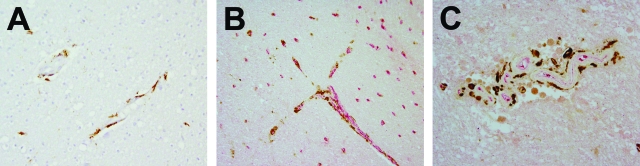
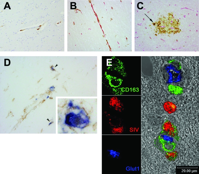
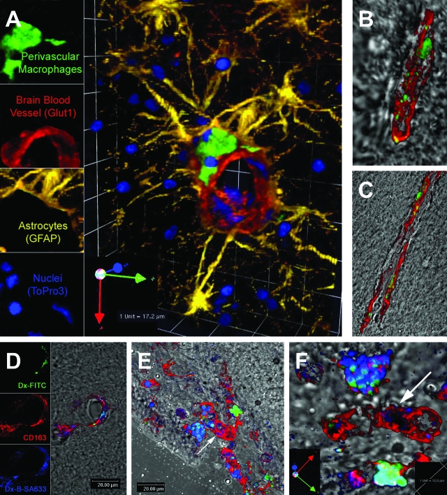
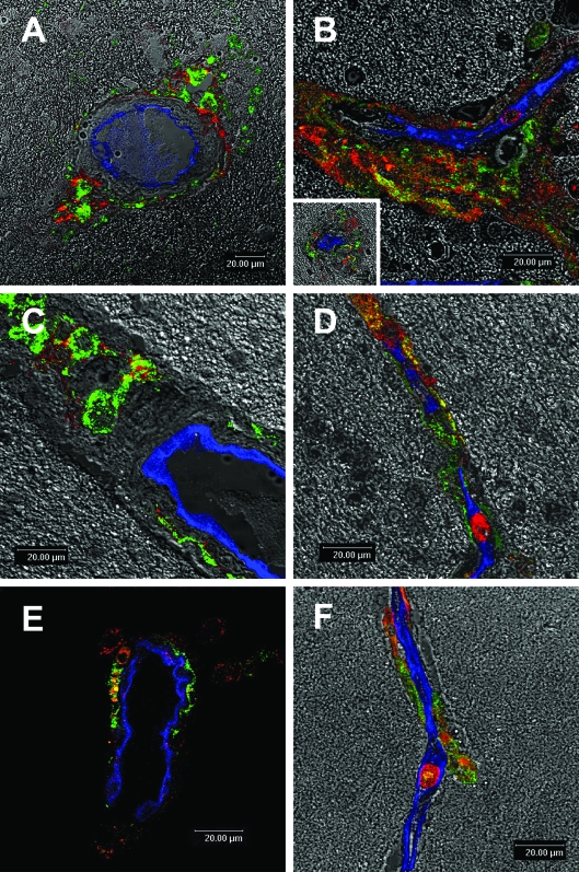
Perivascular macrophages are uniquely situated at the intersection between the nervous and immune systems. Although combined myeloid marker detection differentiates perivascular from resident brain macrophages (parenchymal microglia), no single marker distinguishes perivascular macrophages in humans and mice. Here, we present the macrophage scavenger receptor CD163 as a marker for perivascular macrophages in humans, monkeys, and mice. CD163 was primarily confined to perivascular macrophages and populations of meningeal and choroid plexus macrophages in normal brains and in brains of humans and monkeys with human immunodeficiency virus or simian immunodeficiency virus (SIV) encephalitis. Scattered microglia in SIV encephalitis lesions and multinucleated giant cells were also CD163 positive. Consistent with prior findings that perivascular macrophages are primary targets of human immunodeficiency virus and SIV, all SIV-infected cells in the brain were CD163 positive. Using fluorescent dyes that definitively and selectively label perivascular macrophages in vivo, we confirmed that dye-labeled simian perivascular macrophages were CD163 positive and able to repopulate the central nervous system within 24 hours. Flow cytometric studies demonstrated a subset of monocytes (CD163(+)CD14(+)CD16(+)) that were immunophenotypically similar to brain perivascular macrophages. These findings recognize CD163(+) blood monocytes/macrophages as a source of brain perivascular macrophages and underscore the utility of this molecule in studying the biology of perivascular macrophages and their precursors in humans, monkeys, and mice.
Figures
Similar articles
-
CD163, a marker of perivascular macrophages, is up-regulated by microglia in simian immunodeficiency virus encephalitis after haptoglobin-hemoglobin complex stimulation and is suggestive of breakdown of the blood-brain barrier.Am J Pathol. 2008 Mar;172(3):725-37. doi: 10.2353/ajpath.2008.070848. Epub 2008 Feb 14. Am J Pathol. 2008. PMID: 18276779 Free PMC article.
-
Monocyte/macrophage trafficking in acquired immunodeficiency syndrome encephalitis: lessons from human and nonhuman primate studies.J Neurovirol. 2008 Aug;14(4):318-26. doi: 10.1080/13550280802132857. J Neurovirol. 2008. PMID: 18780233 Free PMC article. Review.
-
A method for obtaining simian immunodeficiency virus RNA sequences from laser capture microdissected and immune captured CD68+ and CD163+ macrophages from frozen tissue sections of bone marrow and brain.J Immunol Methods. 2017 Mar;442:59-63. doi: 10.1016/j.jim.2017.01.003. Epub 2017 Jan 14. J Immunol Methods. 2017. PMID: 28093272 Free PMC article.
-
CD163/CD16 coexpression by circulating monocytes/macrophages in HIV: potential biomarkers for HIV infection and AIDS progression.AIDS Res Hum Retroviruses. 2008 Mar;24(3):417-21. doi: 10.1089/aid.2007.0193. AIDS Res Hum Retroviruses. 2008. PMID: 18373432 Free PMC article.
-
The role of monocytes and perivascular macrophages in HIV and SIV neuropathogenesis: information from non-human primate models.Neurotox Res. 2005 Oct;8(1-2):107-15. doi: 10.1007/BF03033823. Neurotox Res. 2005. PMID: 16260389 Review.
Cited by
-
SIV encephalitis lesions are composed of CD163(+) macrophages present in the central nervous system during early SIV infection and SIV-positive macrophages recruited terminally with AIDS.Am J Pathol. 2015 Jun;185(6):1649-65. doi: 10.1016/j.ajpath.2015.01.033. Epub 2015 May 8. Am J Pathol. 2015. PMID: 25963554 Free PMC article.
-
Monocyte mobilization, activation markers, and unique macrophage populations in the brain: observations from SIV infected monkeys are informative with regard to pathogenic mechanisms of HIV infection in humans.J Neuroimmune Pharmacol. 2012 Jun;7(2):363-71. doi: 10.1007/s11481-011-9330-3. Epub 2011 Dec 14. J Neuroimmune Pharmacol. 2012. PMID: 22167311 Review.
-
Role for cFMS in maintaining alternative macrophage polarization in SIV infection: implications for HIV neuropathogenesis.J Neuroinflammation. 2015 Mar 25;12:58. doi: 10.1186/s12974-015-0272-1. J Neuroinflammation. 2015. PMID: 25886134 Free PMC article.
-
Mechanisms of HIV entry into the CNS: increased sensitivity of HIV infected CD14+CD16+ monocytes to CCL2 and key roles of CCR2, JAM-A, and ALCAM in diapedesis.PLoS One. 2013 Jul 26;8(7):e69270. doi: 10.1371/journal.pone.0069270. Print 2013. PLoS One. 2013. PMID: 23922698 Free PMC article.
-
Selective targeting of perivascular macrophages for clearance of beta-amyloid in cerebral amyloid angiopathy.Proc Natl Acad Sci U S A. 2009 Jan 27;106(4):1261-6. doi: 10.1073/pnas.0805453106. Epub 2009 Jan 21. Proc Natl Acad Sci U S A. 2009. PMID: 19164591 Free PMC article.
References
-
- Gonzalez-Scarano F, Baltuch G. Microglia as mediators of inflammatory and degenerative diseases. Annu Rev Neurosci. 1999;22:219–240. - PubMed
-
- Stoll G, Jander S. The role of microglia and macrophages in the pathophysiology of the CNS. Prog Neurobiol. 1999;58:233–247. - PubMed
-
- Hickey WF, Kimura H. Perivascular microglial cells of the CNS are bone marrow-derived and present antigen in vivo. Science. 1988;239:290–292. - PubMed
-
- Hickey WF, Vass K, Lassmann H. Bone marrow-derived elements in the central nervous system: an immunohistochemical and ultrastructural survey of rat chimeras. J Neuropathol Exp Neurol. 1992;5:246–256. - PubMed
-
- Lassmann H, Schmied M, Vass K, Hickey WF. Bone marrow derived elements and resident microglia in brain inflammation. Glia. 1993;7:19–24. - PubMed
Publication types
MeSH terms
Substances
Grants and funding
LinkOut - more resources
Full Text Sources
Other Literature Sources
Medical
Research Materials
