. 2006 Mar;168(3):898-906.
doi: 10.2353/ajpath.2006.050350.

Transmission of amyloidosis in offspring of mice with AApoAII amyloidosis

Affiliations

Transmission of amyloidosis in offspring of mice with AApoAII amyloidosis

Tatsumi Korenaga et al. Am J Pathol. 2006 Mar.

Abstract

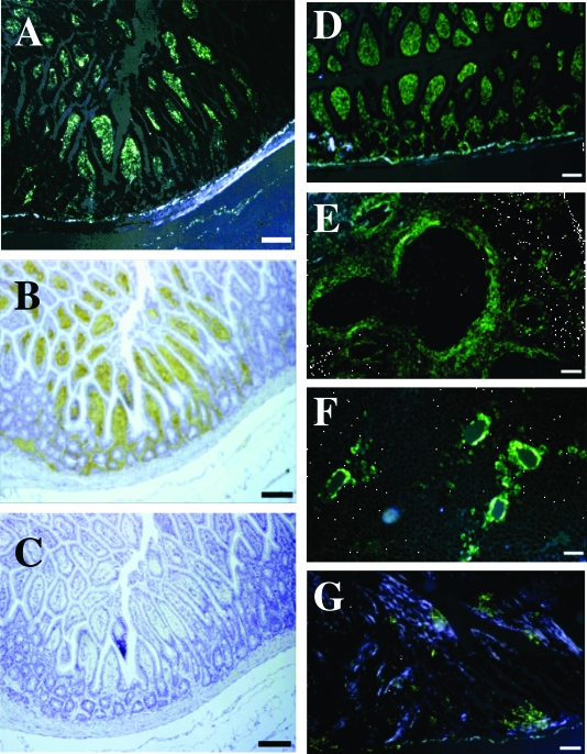
Pre-existing amyloid fibrils can induce further polymerization of endogenous precursor proteins in vivo. Thus, transmission of amyloid fibrils (AApoAII) may induce a conformational change in endogenous apolipoprotein A-II and accelerate amyloid deposition in mouse senile amyloidosis. To characterize transmissibility, we examined amyloidosis in the offspring of AApoAII-injected mother mice that possessed the amyloidogenic Apoa2(c) allele of the apolipoprotein A-II gene. At 4 months of age, amyloid deposits were detected in the intestines of offspring born from and nursed by amyloid fibril-injected mothers, with intensity of deposition increasing thereafter. No amyloid deposits were detected in the offspring of noninjected control mothers. Accelerated amyloidosis was also observed in offspring born from mothers without injection but nursed by amyloid fibril-injected mothers. However, this was not observed in offspring born from amyloid fibril-injected mothers but nursed by control mothers. This fostering excluded vertical transmission through the placenta, suggesting the presence of factors that accelerate amyloidosis during the nursing period. In addition, milk obtained from amyloid fibril-injected mothers induced AApoAII amyloidosis in young mice, and transmission electron microscopy detected noodle-like amyloid fibrils in milk of amyloid fibril-injected mothers. These results provide important insight into the etiology and pathogenesis of amyloid diseases.


Figures

Figure 1
Figure 1
A schematic of events leading from the induction of amyloidosis in parental mice until the determination of amyloid deposition in offspring. Solid and dotted lines represent parents with and without induction of amyloidosis, respectively. Offspring (thinner solid lines) were sacrificed at the ages shown by arrowheads. A: Analysis of maternal transmission. 1, female 2-month-old R1.P1-Apoa2c mice were amyloidosis induced by intravenous injection with AApoAII amyloid fibrils. Control mice were injected with DW. Three months after injection, mice were mated with normal male mice. Offspring were nursed for 21 days, weaned, and sacrificed at 4, 6, 8, and 12 months of age. 2, offspring born from amyloid fibril-injected and control mothers were exchanged just after birth. B: Paternal transmission. Male 2-month-old R1.P1-Apoa2c mice were induced for amyloidosis. Three months after induction, the male mice were mated with normal female mice. The offspring were separated from their parents at the age of 21 days. C: Analysis of induction by milk. Milk of amyloid fibril-injected mother was injected into 1-month-old R1.P1-Apoa2c mice intraperitoneally.
Figure 2
Figure 2
Amyloid deposition in mice born from amyloid fibril-injected mothers. Amyloid deposition was histologically detected by green birefringence in Congo Red-stained sections (A, D, E, F, and G), and amyloid proteins were immunohistochemically identified with anti-apoA-II (B) and anti-AA antiserum (C). A: Amyloid deposit (grade 2) in the intestine of a 4-month-old offspring. B: AApoAII deposits identified in the intestine by staining with anti-apoA-II antiserum. C: No AA was detected in the intestine by staining with anti-AA antiserum. Amyloid deposition in the small intestine (D) (grade 4), spleen (E) (grade 3), liver (F) (grade 3), and heart (G) (grade 2) of 8-month-old offspring. Scale bars = 100 μm.
Figure 3
Figure 3
. Intensity of amyloid deposition in mice born from amyloid fibril-injected mothers. A: Intensity of amyloid deposition was determined in offspring mice of amyloid fibril-injected (•) or control mothers (▵). Litters of 10, 7, and 11 offspring from amyloid fibril-injected mothers and 7, 3, and 9 offspring from control mothers were sacrificed at the ages of 4, 6, and 8 months, respectively. AI was determined from Congo Red-stained sections from seven tissues, plotted versus age. *P < 0.05; **P < 0.01. B: Intensity of amyloid deposition in R1.P1-Apoa2c mice born from control mothers and nursed by amyloid fibril-injected foster mothers (•) and mice born from amyloid fibril-injected mothers and nursed by control foster mothers (○). Ten, 7, and 8 offspring nursed by amyloid fibril-injected mothers and 5, 9, and 5 offspring nursed by control mothers were sacrificed at the age of 4, 6, and 8 months, respectively. AI determined from Congo Red-stained sections from seven tissues was plotted versus age. *P < 0.05; **P < 0.01.
Figure 4
Figure 4
Detection of apoA-II protein in milk and urine. Western blotting analysis of milk and urine from amyloid fibril-injected R1.P1-Apoa2c mice, detected with anti-apoA-II antiserum. A: Lane 1, kaleidoscope prestained standard markers. Lanes 2 and 3, 1 μl of Coomassie Brilliant Blue R 250-stained milk from an amyloid fibril-injected mother (6 months old) and control mother (6 months old), respectively. Lanes 4, 5, 6, 7, 8, 9, and 10, Western blotting analysis of milk reacted with anti-apoA-II antiserum. Lane 4, 1 μl of milk from an amyloid fibril-injected mother; Lane 5, 1 μl of milk from a control mother. Lane 6, 50 μg of proteins from 0.15 mol/L NaCl supernatant centrifuged at 40,000 × g from the milk from an amyloid fibril-injected mother. Lane 7, 20 μg of proteins from the water suspension (amyloid fibril) fraction of milk from an amyloid fibril-injected mother. Lane 8, 50 μg of proteins from the 0.15 mol/L NaCl supernatant of milk from a control mother. Lane 9, 20 μg of proteins from the water suspension fraction of milk from a control mother. Lane 10, AApoAII amyloid fibril. B: Lane 1, 100 μg of proteins from lyophilized whole urine of amyloid fibril-injected mice; lane 2, 100 μg of proteins from lyophilized whole urine of control mice; lane 3, pellets centrifuged at 100,000 × g from 4.0 ml of urine from amyloid fibril-injected mice; lane 4, pellets from urine of control mice; and lane 5, AApoAII amyloid fibril.
Figure 5
Figure 5
Transmission electron microscopy images of amyloid fibrils extracted from milk of an amyloid fibril-injected mother. Scale bar = 100 nm.

Similar articles

Cited by

References

    1. Selkoe DJ. Folding proteins in fatal ways. Nature. 2003;426:900–904. - PubMed
    1. Glenner GG. Amyloid deposits and amyloidosis: the beta-fibrilloses. N Engl J Med. 1980;302:1283–1292. - PubMed
    1. Merlini G, Bellotti V. Molecular mechanisms of amyloidosis. N Engl J Med. 2003;349:583–596. - PubMed
    1. Jarrett JT, Lansbury PT., Jr Seeding “one-dimensional crystallization” of amyloid: a pathogenic mechanism in Alzheimer’s disease and scrapie? Cell. 1993;73:1055–1058. - PubMed
    1. Rochet JC, Lansbury PT., Jr Amyloid fibrillogenesis: themes and variations. Curr Opin Struct Biol. 2000;10:60–68. - PubMed

Publication types