. 2006 Mar;168(3):1004-21.
doi: 10.2353/ajpath.2006.050471.

Identification of differential protein expression associated with development of unstable human carotid plaques

Affiliations

Identification of differential protein expression associated with development of unstable human carotid plaques

Mark Slevin et al. Am J Pathol. 2006 Mar.

Abstract

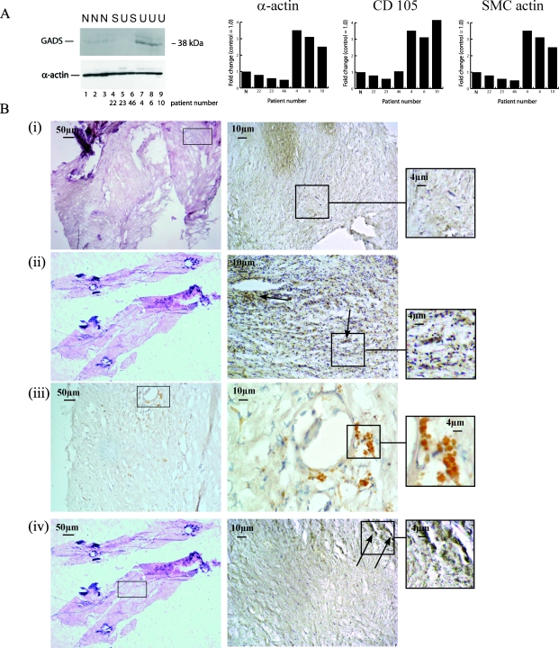
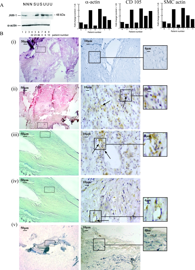
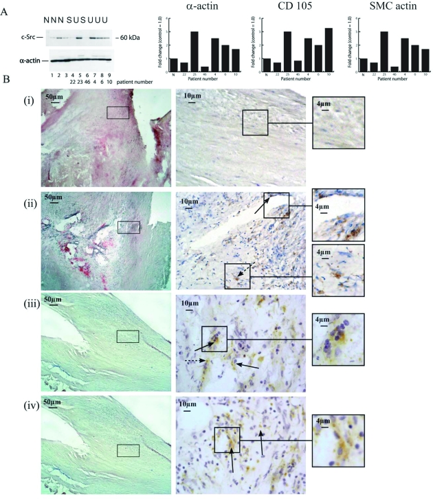
Rupture-prone unstable arterial plaques develop concomitantly with the appearance of intraplaque hemorrhage and tissue ulceration, in association with deregulation of smooth muscle cell mitogenesis and leakage of newly formed blood vessels. Using microarray technology, we have identified novel protein deregulation associated with unstable carotid plaque regions. Overexpression of proapoptotic proteins caspase-9 and TRAF4 was seen in endothelial cells and smooth muscle cells from unstable hemorrhagic and ulcerated plaque regions. Topoisomerase-II-alpha (TOPO-II-alpha), which is associated with DNA repair mechanisms, was also overexpressed by these cells. Cell signaling molecules c-src, G-protein-coupled receptor kinase-interacting protein (GIT1), and c-jun N-terminal kinase (JNK) were up-regulated in endothelial cells from the same areas, whereas an increase in expression of junctional adhesion molecule-1 (JAM-1) in blood vessels and infiltrating macrophages from inflammatory regions might form part of a leukocyte rolling response, increasing the plaque volume. Grb2-like adaptor protein (Gads), responsible for differentiation of monocytes into macrophages, was expressed by macrophages from unstable plaques, suggesting a potential mechanism through which increased scavenging could occur in rupture-prone areas. We conclude that modulation of novel cell signaling intermediates, such as those described here, could be useful in the therapy of angiogenesis and apoptosis, designed to reduce unstable plaque formation.


Figures

Figure 1
Figure 1
A: Western blot showing caspase-9 (46 kd) and cleaved caspase-9 (34 kd) expression in normal arteries obtained at transplant (lanes 1–3); stable, fibrous arteries (lanes 4 and 6); and unstable hemorrhagic/ulcerated arteries (lanes 5 and 7–9). Increased expression was seen in the latter. In the bar charts, left represents a comparison against α-actin (loading control), right is with SMC actin, and middle versus EC content (anti-CD105). The mean value for the three normal arteries is presented as N. B: Immunohistochemical localization of caspase-9 in fibrotic portions of artery showing weak staining (i, patient 58), and in unstable plaques, showing overexpression in macrophages (ii, arrow; patient 52), SMCs (iii, arrow; patient 3), and ECs (iv, arrows; patient 3) from hemorrhagic regions. v is a double-labeled section showing caspase-9 association with macrophages (patient 78). Macrophages (anti-CD68) were stained with Vector blue. Staining was performed using the VectaStain ABC kit. Original magnifications (B, i–v): ×10 (left); ×40 (middle); and ×100 (right).
Figure 2
Figure 2
A: Western blot showing GADS (34 kd) expression in normal arteries obtained at transplant (lanes 1–3); stable, fibrous arteries (lanes 4 and 6); and unstable hemorrhagic/ulcerated arteries (lanes 5 and 7–9). Increased expression was seen in the latter. In the bar charts, left represents a comparison against α-actin (loading control), right is with SMC actin, and middle versus EC content (anti-CD105). The mean value for the three normal arteries is presented as N. B: Immunohistochemical localization of GADS showed weak staining in fibrous regions (i, patient 29), increased expression in macrophages from unstable regions (ii and iii, arrows; patients 29 and 65, respectively), and association with ECs from hemorrhagic regions (iv, arrows; patient 29). Staining was performed using the VectaStain ABC kit. Original magnifications (B, i–iv): ×10 (left); ×40 (middle); and ×100 (right).
Figure 3
Figure 3
A: Western blot showing expression of GIT1 (95 kd) expression in normal arteries obtained at transplant (lanes 1–3); stable, fibrous arteries (lanes 4–6); and unstable hemorrhagic/ulcerated arteries (lanes 7–9). Increased expression was seen in lanes 7–9. In the bar charts, left represents a comparison against α-actin (loading control), right is with SMC actin, and middle versus EC content (anti-CD105). The mean value for the three normal arteries is presented as N. B: Immunohistochemical localization demonstrated weak staining in normal looking and fibrous regions (i, patient 58) but strong staining in infiltrating macrophages from ulcerated unstable plaques with high cellular content (ii, arrows; patient 3; iii, patient 52; and iv, patient 28). v is a double-labeled section showing GIT1 association with macrophages (patient 65). Macrophages (anti-CD68) were stained with Vector blue. Staining was performed using the VectaStain ABC kit. Original magnifications (B, i–v): ×10 (left); ×40 (middle); and ×100 (right).
Figure 4
Figure 4
A: Western blot showing expression of JAM-1 (48 kd) protein in normal arteries obtained at transplant (lanes 1–3); stable, fibrous arteries (lanes 4 and 6); and unstable hemorrhagic/ulcerated arteries (lanes 5 and 7–9). Increased expression can be seen in unstable arteries. In the bar charts, left represents a comparison against α-actin (loading control), right is with SMC actin, and middle versus EC content (anti-CD105). The mean value for the three normal arteries is presented as N. B: Immunohistochemistry showed weak staining in fibrotic areas (i, patient 29) and increased expression in macrophages (ii, arrows; patient 40) and ECs (iii and iv, arrows; patient 54) from unstable hemorrhagic and ulcerated regions. v is a double-labeled section showing JAM-1 association with ECs (patient 65). ECs (anti-CD105) were stained with Vector blue. Staining was performed using the VectaStain ABC kit. Original magnifications (B, i–v): ×10 (left); ×40 (middle); and ×100 (right).
Figure 5
Figure 5
A: Western blot shows expression of JNK (JNK1, 43 kd; JNK2, 51 kd) in normal arteries obtained at transplant (lanes 1–3); stable, fibrous arteries (lanes 4–6); and unstable hemorrhagic/ulcerated arteries (lanes 7–9). Increased expression of both JNK1 and JNK2 was seen in stable and unstable plaques (lanes 4–9) compared with control arteries (lanes 1–3). In the bar charts, left represents a comparison against α-actin (loading control), right is with SMC actin, and middle versus EC content (anti-CD105). The mean value for the three normal arteries is presented as N. B: Immunohistochemistry showed weak staining of SMCs in fibrous arteries (i, patient 121) but was strongly expressed by macrophages (ii, arrows; patient 65) and SMCs (iii, arrows; patient 65) in ulcerated and hemorrhagic plaques. Staining was performed using the VectaStain ABC kit. Original magnifications (B, i–iii): ×10 (left); ×40 (middle); and ×100 (right).
Figure 6
Figure 6
A: Western blot shows expression of c-src in normal arteries obtained at transplant (lanes 1–3); stable, fibrous arteries (lanes 4 and 6); and unstable hemorrhagic/ulcerated arteries (lanes 5 and 7–9). Increased expression of c-src was seen particularly in unstable plaques (lanes 5 and 7–9) compared with control arteries. In the bar charts, left represents a comparison against α-actin (loading control), right is with SMC actin, and middle versus EC content (anti-CD105). The mean value for the three normal arteries is presented as N. B: Immunohistochemistry demonstrated weak staining of c-src in fibrous regions (i, patient 28), and increased immunoreactivity associated with ECs (arrows) and macrophages (broken arrows) from unstable plaque regions (ii, arrows; patient 52; iii and iv, arrows; patient 54). Staining was performed using the VectaStain ABC kit. Original magnifications (B, i–iv): ×10 (left); ×40 (middle); and ×100 (right).
Figure 7
Figure 7
A: Western blot shows expression of native Topo-II-α (170 kd) in normal arteries obtained at transplant (lanes 1–3); stable, fibrous arteries (lanes 4–6); and unstable hemorrhagic/ulcerated arteries (lanes 7–9). Increased expression of Topo-II-α was seen in the majority of plaques, and the presence of the cleaved protein (70 kd) can be seen in lanes 6 and 7. In the bar charts, left represents a comparison against α-actin (loading control), right is with SMC actin, and middle versus EC content (anti-CD105). The mean value for the three normal arteries is presented as N. B: Immunohistochemistry showed weak staining in fibrous plaques (i, patient 28) and increased immunoreactivity in ulcerated plaques, particularly associated with ECs (arrows) and SMCs (broken arrows) (ii and iii, arrows; patient 28). iv is a double-labeled section showing Topo-II-α association with SMCs (patient 78, arrows). SMCs (anti-SMC-actin) were stained with Vector blue. Staining was performed using the VectaStain ABC kit. Original magnifications (B, i–v): ×10 (left); ×40 (middle); and ×100 (right).
Figure 8
Figure 8
A: Western blot shows expression of TRAF4 in normal arteries obtained at transplant (lanes 1–3); stable, fibrous arteries (lanes 4 and 6); and unstable hemorrhagic/ulcerated arteries (lanes 5 and 7–9). Increased expression of TRAF4 was seen in unstable plaques compared with control arteries and stable plaques. In the bar charts, left represents a comparison against α-actin (loading control), right is with SMC actin, and middle versus EC content (anti-CD105). The mean value for the three normal arteries is presented as N. B: Immunohistochemistry showed weak staining of TRAF4 in fibrotic areas of arteries (i, patient 29) but strong expression in blood vessels (arrows) and infiltrating lymphocytes and macrophages (broken arrows) from unstable hemorrhagic plaques (ii and iii, arrows; patient 40; iv, arrows; patient 54). v is a double-labeled section showing TRAF4 association with ECs (patient 78, arrows). ECs (anti-CD105) were stained with Vector blue. Staining was performed using the VectaStain ABC kit. Original magnifications (B, i–v): ×10 (left); ×40 (middle); and ×100 (right).

References

    1. Hennerici MG. The unstable plaque. Cerebrovasc Dis. 2004;17:17–22. - PubMed
    1. Moulton KS. Plaque angiogenesis: its function and regulation. Cold Spring Harbor Symp Quant Biol. 2002;11:471–482. - PubMed
    1. DeGraba TJ, Siren AL, Penix L, McCarron RM, Hargraves R, Sood S, Pettigrew KD, Hallenbeck JM. Increased endothelial expression of intercellular adhesion molecule-1 in symptomatic versus asymptomatic human carotid atherosclerotic plaque. Stroke. 1998;29:1405–1410. - PubMed
    1. Loftus IM, Naylor AR, Goodall S, Crowther M, Jones L, Bell PR, Thompson MM. Increased matrix metalloproteinase-9 activity in unstable carotid plaques. A potential role in acute plaque disruption. Stroke. 2000;31:40–47. - PubMed
    1. Mallat Z, Corbaz A, Scoazec A, Besnard S, Leseche G, Chvatchko Y, Tedgui A. Expression of interleukin-18 in human atherosclerotic plaques and relation to plaque instability. Circulation. 2001;104:1598–1603. - PubMed