. 2006 Mar;168(3):1022-30.
doi: 10.2353/ajpath.2006.050868.

Renewal of mural thrombus releases plasma markers and is involved in aortic abdominal aneurysm evolution

Affiliations

Renewal of mural thrombus releases plasma markers and is involved in aortic abdominal aneurysm evolution

Ziad Touat et al. Am J Pathol. 2006 Mar.

Abstract

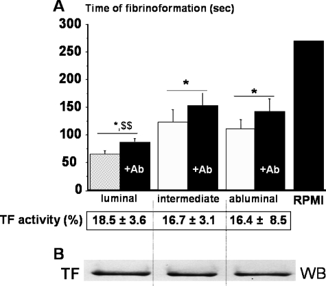
Human abdominal aortic aneurysm (AAA) expansion has been linked to the presence of a mural thrombus. Here we explored the mechanism of the continual luminal renewal of this thrombus and its ability to release biological markers potentially detectable in plasma. We also explored the ability of platelet inhibition to pacify the thrombus and to limit aneurysm progression in an experimental model. Blood samples and mural thrombi were collected in 20 AAA patients. In parallel, segments of sodium dodecyl sulfate-decellularized guinea pig aorta were xenografted onto the abdominal aorta of 30 rats to induce aneurysms. Fifteen rats received abciximab treatment and fifteen received irrelevant immunoglobulins. Procoagulant activity and platelet activation markers (microparticles, sP-selectin, sGPV, sCD40L) were increased threefold to fivefold in eluates from the luminal thrombus layer compared to other layers. All these markers were increased twofold to fivefold in patients' plasma compared to matched controls (P < 0.005). In the rat model, abciximab reduced both thrombus area and aneurysmal enlargement (P < 0.05). Platelet aggregation is probably responsible for the renewal of the thrombus in AAA. The luminal thrombus released markers of platelet activation that could easily be detected in plasma. Platelet inhibition limited aortic aneurysm expansion in a rat model, providing new therapeutic perspectives in the prevention of AAA enlargement.


Figures

Figure 1
Figure 1
Topography of platelet activation in the luminal layer of the thrombus. A: Macroscopic aspect of the mural thrombus showing the three layers: fresh red luminal layer, compact brown intermediate layer, and granular fibrinolytic abluminal layer. B: Microscopic aspect (Masson’s trichrome, n = 8) showing fibrin (gray) and red blood cells (red) in the luminal layer, more compact fibrin polymer including broken-down red blood cells in the intermediate layer and the loose mesh of degraded fibrin in the abluminal layer. C: Cell nuclear staining by 4′,6′-diamidino-2-phenylindole hydrochloride (n = 6). D: DNA fragmentation visualized by the terminal dUTP nick-end labeling method (n = 6). E: Immunostaining of GPIIIa (CD61, β3) predominated in the luminal layer, reproducing the luminal patchy network: brown areas, arrows corresponding to GPIIIa/fibrin-rich areas, gray areas corresponding to red blood cell accumulation (top, n = 4). These areas were absent in the abluminal layer (bottom). F: Immunostaining of P-selectin (CD62P) also predominated in the luminal layer corresponding to GPIIIa/fibrin-rich areas (brown areas, arrows, top; n = 4). Trapped PMNs were co-localized with P-selectin in fibrin-rich areas (hematoxylin-stained nuclei, bottom). Original magnifications: ×100 (B–D); ×400 [E, F (top)]; ×1000 (F, bottom).
Figure 2
Figure 2
Role of TF in fibrin generation. A: Time of fibrin generation of citrated plasma (clotting time) in presence of CaCl2 and eluates from the different layers with (filled bar) and without (open bar) blocking TF antibody (RPMI used as control, n = 18). Time of fibrin formation was significantly shorter in luminal eluates as compared to those of intermediary and abluminal layers ($$P < 0.005). For each layer, TF antibody increased significantly (*P < 0.05) the clotting time by 18.5, 16.7, and 16.4%, respectively. This TF-dependent increase was not significantly different between the three layers. B: Moreover, a homogeneous distribution of TF throughout the thrombus layers was shown by Western blot (WB, n = 6).
Figure 3
Figure 3
Luminal thrombus released more phospholipids than other layers. A: Phosphatidylserine staining by biotinylated annexin V predominated in the luminal layer (n = 4). B: Chromogenic assays of TF (pg/g) and phosphatidylserine (pmol/L/g) procoagulant activities. There was no significant difference for TF in the three layers. In contrast, phosphatidylserine is more exposed in the luminal layer as compared to intermediate and abluminal layers (P < 0.005, n = 12). Original magnifications, ×200.
Figure 4-6770
Figure 4-6770
Blocking α2bβ3 promoted cell recolonization of the thrombus and limited aneurysm expansion in an experimental model of aneurysm in rats. A: Macroscopic aspects of experimental aneurysm (Sirius red staining) showed the important enlargement and thrombus (T) in controls (n = 15) as compared to abciximab-treated rats (n = 15) (less enlargement and small thrombus). B: P-selectin immunostaining predominated in the luminal thrombus layer of controls, whereas it was absent in abciximab-treated rats. C: H&E staining showed the luminal accumulation of PMNs in control thrombi (arrow) whereas abciximab limited this infiltration in the luminal layer of the thrombus. D: Elastic lamellae (arrows) were completely disorganized in thrombus-exposed media of controls whereas elastin was partly preserved in abciximab-treated rats. E: α-Actin-positive cells (arrows) were rarely present in the luminal layer of the thrombus in the control group. Moreover, these rare mesenchymatous cells did not spread on the fibrin (round morphology) and underwent apoptosis as suggested by the presence of pycnotic nuclei (top inset). In contrast, α-actin-positive cells appeared to spread and proliferate in the thrombus of abciximab-treated rats. Inhibition of platelet aggregation lead to limitation of thrombus area (F) and aneurysm diameter (G) in treated rats (*P < 0.05, n = 15). H: Lastly, there was a correlation between thrombus area and aneurysm dilatation in each group (controls, +; abciximab group, filled circle) that differed by the slope (P < 0.05). Original magnifications: ×2 (A); ×400 (B–E).

Similar articles

Cited by

References

    1. Brady AR, Thompson SG, Fowkes FG, Greenhalgh RM, Powell JT. Abdominal aortic aneurysm expansion: risk factors and time intervals for surveillance. Circulation. 2004;110:16–21. - PubMed
    1. Sakalihasan N, Limet R, Defawe OD. Abdominal aortic aneurysm. Lancet. 2005;365:1577–1589. - PubMed
    1. Fontaine V, Jacob MP, Houard X, Rossignol P, Plissonnier D, Angles-Cano E, Michel JB. Involvement of the mural thrombus as a site of protease release and activation in human aortic aneurysms. Am J Pathol. 2002;161:1701–1710. - PMC - PubMed
    1. Fontaine V, Touat Z, Mtairag el M, Vranckx R, Louedec L, Houard X, Andreassian B, Sebbag U, Palombi T, Jacob MP, Meilhac O, Michel JB. Role of leukocyte elastase in preventing cellular re-colonization of the mural thrombus. Am J Pathol. 2004;164:2077–2087. - PMC - PubMed
    1. Adolph R, Vorp DA, Steed DL, Webster MW, Kameneva MV, Watkins SC. Cellular content and permeability of intraluminal thrombus in abdominal aortic aneurysm. J Vasc Surg. 1997;25:916–926. - PubMed

Publication types

MeSH terms