

The sialomucin CD34 is a marker of lymphatic endothelial cells in human tumors
- PMID: 16507917
- PMCID: PMC1606520
- DOI: 10.2353/ajpath.2006.050554
The sialomucin CD34 is a marker of lymphatic endothelial cells in human tumors
Abstract
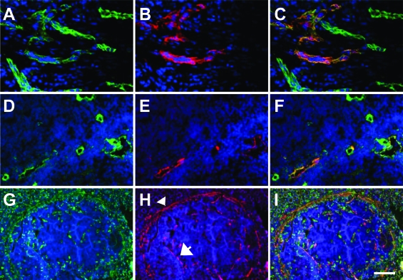
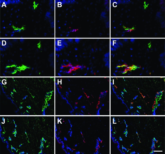
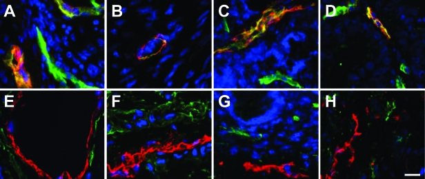
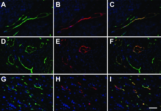
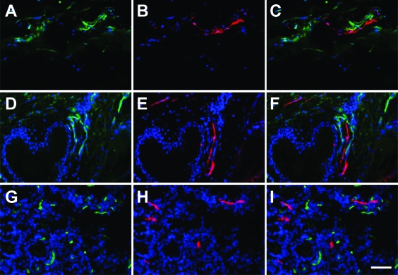
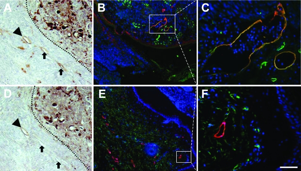
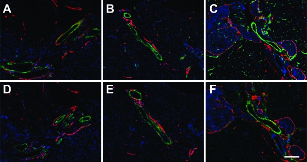
The mechanisms of lymphangiogenesis have been increasingly understood in recent years. Yet, the contribution of lymphangiogenesis versus lymphatic cooption in human tumors and the functionality of tumor lymphatics are still controversial. Furthermore, despite the identification of lymphatic endothelial cell (LEC) markers such as Prox1, podoplanin, LYVE-1, and VEGFR-3, no activation marker for tumor-associated LECs has been identified. Applying double-staining techniques with established LEC markers, we have screened endothelial cell differentiation antigens for their expression in LECs. These experiments identified the sialomucin CD34 as being exclusively expressed by LECs in human tumors but not in corresponding normal tissues. CD34 is expressed by LYVE-1(+)/podoplanin(+)/Prox1(+) tumor-associated LECs in colon, breast, lung, and skin tumors. More than 60% of analyzed tumors contained detectable intratumoral lymphatics. Of these, more than 80% showed complete co-localization of CD34 with LEC markers. In contrast, LECs in all analyzed normal organs did not express CD34. Corresponding analyses of experimental tumors revealed that mouse tumor-associated LECs do not express CD34. Taken together, these experiments identify CD34 as the first differentially expressed LEC antigen that is selectively expressed by tumor-associated LECs. The data warrant further exploration of CD34 in tumor-associated LECs as a prognostic tumor marker.
Figures
Similar articles
-
The transcription factor Prox1 is a marker for lymphatic endothelial cells in normal and diseased human tissues.FASEB J. 2002 Aug;16(10):1271-3. doi: 10.1096/fj.01-1010fje. Epub 2002 Jun 7. FASEB J. 2002. PMID: 12060670
-
Lymphatic endothelial cells, tumor lymphangiogenesis and metastasis: New insights into intratumoral and peritumoral lymphatics.Cancer Metastasis Rev. 2006 Dec;25(4):677-94. doi: 10.1007/s10555-006-9026-y. Cancer Metastasis Rev. 2006. PMID: 17160713 Review.
-
The role of podoplanin in tumor progression and metastasis.Anticancer Res. 2008 Sep-Oct;28(5B):2997-3006. Anticancer Res. 2008. PMID: 19031946 Review.
-
Isolation and characterization of lymphatic endothelial cells from human glossal lymphangioma.Oncol Rep. 2010 Jan;23(1):105-11. Oncol Rep. 2010. PMID: 19956870
-
Elevated expression of VEGFR-3 in lymphatic endothelial cells from lymphangiomas.BMC Cancer. 2007 Jun 21;7:105. doi: 10.1186/1471-2407-7-105. BMC Cancer. 2007. PMID: 17584927 Free PMC article.
Cited by
-
Lymphatic metastasis in breast cancer: importance and new insights into cellular and molecular mechanisms.Clin Exp Metastasis. 2007;24(8):619-36. doi: 10.1007/s10585-007-9123-5. Epub 2007 Nov 6. Clin Exp Metastasis. 2007. PMID: 17985200 Review.
-
Radiogenic lymphangiogenesis in the skin.Am J Pathol. 2007 Jul;171(1):338-48. doi: 10.2353/ajpath.2007.060589. Am J Pathol. 2007. PMID: 17591978 Free PMC article.
-
A dual 5α-reductase inhibitor dutasteride caused reductions in vascular density and area in benign prostatic hyperplasia.ISRN Urol. 2013;2013:863489. doi: 10.1155/2013/863489. Epub 2013 Jan 17. ISRN Urol. 2013. PMID: 23401800 Free PMC article.
-
Expression and clinical implication of cyclooxygenase-2 and E-cadherin in oral squamous cell carcinomas.Cancer Biol Ther. 2020 Aug 2;21(8):667-674. doi: 10.1080/15384047.2015.1071741. Epub 2020 Jun 17. Cancer Biol Ther. 2020. PMID: 26218314 Free PMC article.
-
Role of tumor-associated lymphatic endothelial cells in metastasis: a study of epithelial ovarian tumor in vitro.Cancer Sci. 2010 Mar;101(3):679-85. doi: 10.1111/j.1349-7006.2009.01436.x. Epub 2009 Nov 14. Cancer Sci. 2010. PMID: 20028387 Free PMC article.
References
-
- Saharinen P, Tammela T, Karkkainen MJ, Alitalo K. Lymphatic vasculature: development, molecular regulation and role in tumor metastasis and inflammation. Trends Immunol. 2004;25:387–395. - PubMed
-
- Breiteneder-Geleff S, Soleiman A, Kowalski H, Horvat R, Amann G, Kriehuber E, Diem K, Weninger W, Tschachler E, Alitalo K, Kerjaschki D. Angiosarcomas express mixed endothelial phenotypes of blood and lymphatic capillaries: podoplanin as a specific marker for lymphatic endothelium. Am J Pathol. 1999;154:385–394. - PMC - PubMed
Publication types
MeSH terms
Substances
LinkOut - more resources
Full Text Sources
Miscellaneous
