. 2006 Mar;26(6):2019-28.
doi: 10.1128/MCB.26.6.2019-2028.2006.

Histone deacetylase inhibitors induce VHL and ubiquitin-independent proteasomal degradation of hypoxia-inducible factor 1alpha

Affiliations

Histone deacetylase inhibitors induce VHL and ubiquitin-independent proteasomal degradation of hypoxia-inducible factor 1alpha

Xianguo Kong et al. Mol Cell Biol. 2006 Mar.

Abstract

Adaptation to hypoxic microenvironment is critical for tumor survival and metastatic spread. Hypoxia-inducible factor 1alpha (HIF-1alpha) plays a key role in this adaptation by stimulating the production of proangiogenic factors and inducing enzymes necessary for anaerobic metabolism. Histone deacetylase inhibitors (HDACIs) produce a marked inhibition of HIF-1alpha expression and are currently in clinical trials partly based on their potent antiangiogenic effects. Although it has been postulated that HDACIs affect HIF-1alpha expression by enhancing its interactions with VHL (von Hippel Lindau), thus promoting its ubiquitination and degradation, the actual mechanisms by which HDACIs decrease HIF-1alpha levels are not clear. Here, we present data indicating that HDACIs induce the proteasomal degradation of HIF-1alpha by a mechanism that is independent of VHL and p53 and does not require the ubiquitin system. This degradation pathway involves the enhanced interaction of HIF-1alpha with HSP70 and is secondary to a disruption of the HSP70/HSP90 axis function that appears mediated by the activity of HDAC-6.


Figures

FIG. 1.
FIG. 1.
TSA represses HIF-1α induction in response to hypoxia. A. Dose responses. U-87 cells were exposed to hypoxia (1% O2) for 8 h in the presence of increasing concentrations of TSA. Cell lysates (40 μg) were analyzed by Western blotting using anti-HIF-1α and tubulin (Tub) monoclonal antibodies. B. Time course. U-87 cells were exposed to 300 nM of TSA and 1% O2 for 4 or 8 h, and HIF-1α levels were analyzed by Western blotting. C. Tumor cell lines. Different cell lines were exposed to dimethyl sulfoxide or 300 nM of TSA for 8 h under 1% O2. D. Effect of TSA on gene expression. Total RNA from U-87 cells exposed to dimethyl sulfoxide or TSA for 8 h under conditions of 21% or 1% O2 was analyzed by RT-PCR using primers described in Materials and Methods. VEGF, vascular endothelial growth factor.
FIG. 2.
FIG. 2.
TSA represses HIF-1α independently of VHL and p53. A. VHL independence. RCC4-VHL+ and -VHL cells were treated under normoxia and hypoxia conditions for 8 h in the presence or absence of TSA (300 nM), and cell lysates were analyzed by Western blotting. Tub, tubulin. B. Dose responses and time courses. Normoxic VHL cells were exposed to increasing concentrations of TSA for 8 h (upper panels) or to 300 nM of TSA for different time periods (lower panels). C. Effect of SAHA on HIF-1α expression. VHL cells were exposed to increasing concentrations of SAHA for 8 h. D. TSA suppresses gene expression in VHL cells. VHL cells were exposed to TSA for 8 h, and total RNA was analyzed by RT-PCR. E. p53 independence. HCT116 p53+ or p53 cells were exposed to TSA (300 nM) for 8 h, and cell lysates were analyzed for HIF-1α and p53 expression. VEGF, vascular endothelial growth factor.
FIG. 3.
FIG. 3.
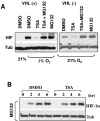
TSA-induced degradation of HIF-1α is mediated by the proteasome system. A. Proteasome inhibitors reverse TSA effect. RCC4 VHL+ or VHL cells were exposed to TSA (300 nM) in the presence or absence of MG132 (5 μM) for 8 h, and cell lysates were analyzed by Western blotting. DMSO, dimethyl sulfoxide. B. TSA does not affect HIF-1α translation. Normoxic U-87 cells were exposed to MG132 (5 μM) in the presence or absence of TSA, and HIF-1α accumulation was measured at 2, 4, and 6 h. Tub, tubulin.
FIG. 4.
FIG. 4.
TSA-induced HIF-1α degradation is independent of ubiquitination. A. TSA degrades HIF-1α in E1-deficient cells. Normoxic Ts20 cells, expressing a temperature-sensitive E1 enzyme, were exposed to 39°C for 8 h in the presence or absence of TSA and in the presence of the proteasome inhibitor MG132 (5 μM) or the indicated concentrations of epoxomicin (Epox), the caspase inhibitor Z-VAD-FMK (50 μM), or the calpain inhibitor Z-Leu-Leu-Cho (40 μM). HIF-1α expression was analyzed by Western blotting. Tub, tubulin. B. E1-competent Ts20 cells respond to TSA. Ts20 cells cultured at 35°C were exposed to hypoxia (1% O2) in the presence of TSA and the inhibitors described for panel A. C. 17AAG degrades HIF-1α in Ts20 cells. Normoxic Ts20 cells were exposed to 39°C for 8 h in the presence or absence of 17AAG (1 μM) and in the presence or absence of the proteasome inhibitor MG132 (5 μM).
FIG. 5.
FIG. 5.
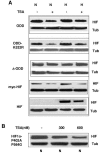
HIF-1α-ODD is sufficient and necessary for TSA-induced degradation. A. ODD determines response to TSA independently of K532 acetylation. HT1080 cells were transfected with plasmids containing the ODD (top pair of panels), a K532R ODD mutant (second pair of panels), an ODD-deleted HIF-1α construct (ΔODD; third pair of panels), or a whole HIF-1α construct (myc-HIF; fourth pair of panels), and the cells were exposed to TSA (300 nM) for 8 h under normoxic (N) or hypoxic (H) conditions. The fifth pair of panels (HIF) presents the endogenous HIF-1α protein. B. The TSA effect is independent of prolyl hydroxylation. A HIF-1α construct containing P402A and P564G mutations was transfected into HT1080 cells and exposed to increasing concentrations of TSA for 8 h under normoxia conditions. Cell lysates were analyzed by Western blotting.
FIG. 6.
FIG. 6.
TSA enhances the interaction between HIF-1α and HSP70. A. HIF-1α interacts with HSP70. RCC4-VHL cells were treated with increasing concentrations of TSA for 4 h, and cell lysates were immunoprecipitated (IP) with anti-HIF-1α monoclonal antibodies. The precipitate was immunoblotted with anti-HIF-1α and anti-HSP70. B. HSP70 interacts with HIF-1a. Cells were treated as described for panel A, but lysates were precipitated with anti-HSP-70 monoclonal antibodies. C. TSA treatment increases HIF-1α in lysate pellets. VHL cells were treated for 6 h in the presence or absence of MG132 and TSA. The cell lysates were centrifuged, and the cell pellets were resuspended in loading buffer and analyzed by Western blotting. D. TSA treatment enhances HSP90 acetylation. RCC4-VHL cells were treated with TSA (600 nM) for 6 h followed by immunoprecipitation with anti-HSP90 antibodies. Whole-cell lysates and immunoprecipitation results were analyzed by Western blotting utilizing anti-HSP90 and anti-acetylated lysine (a-AcLys) antibodies. E. TSA treatment decreases the interaction between HSP90 and HIF-1α. RCC4-VHL cells were incubated with TSA (600 nM) for 6 h followed by immunoprecipitation with anti-HIF-1α antibodies. The immunoprecipitation results was analyzed by Western blotting using anti-HIF-1α and anti-HSP90 antibodies.
FIG. 7.
FIG. 7.
HDAC-6 is involved in the TSA-induced degradation of HIF-1α. A. Overexpression of HDAC-6 enhances HIF-1α levels in response to hypoxia. Control A549 (vector) and overexpressing HDAC-6 cells were subjected to 1% O2 for 8 h at the indicated concentrations of TSA. Cell lysates were analyzed by Western blotting using anti-HIF-1α, anti-HDAC-6, and anti-tubulin (Tub) antibodies. B. Knockdown of HDAC-6 reduces HIF-1α levels in response to hypoxia. Control 293T cells (Vector) and HDAC-6 knockdown cells (KD) were treated and analyzed by Western blotting as described for panel A. C. Knockdown of HDAC-6 suppresses hypoxia-inducible gene expression. Vector and knockdown cells were treated as described for panel A, and total RNA was analyzed by RT-PCR. VEGF, vascular endothelial growth factor.
FIG. 8.
FIG. 8.
Schematic representation of the mechanisms involved in HIF-1α degradation following HDACI treatment. A. In the absence of HDACIs, newly synthesized HIF-1α molecules interact with its chaperones HSP70 and HSP90 to complete its maturation. Under normoxic conditions (+O2), the mature protein is hydroxylated, ubiquitinated, and degraded by the 26S proteasome, while under hypoxia conditions (−O2), the protein survives, interacts with ARNT, and binds hypoxia response element (HRE) sequences to initiate transcription. B. During HDACI treatment, HDAC-6 inhibition results in hyperacetylation of HSP90, accumulation of immature HIF-1α protein/HSP70 complex, and degradation of HIF-1α by the 20S proteasome.

Similar articles

Cited by

References

    1. Arnesen, T., D. Anderson, C. Baldersheim, M. Lanotte, J. E. Varhaug, and J. R. Lillehaug. 2004. Identification and characterization of the human ARD1-NATH protein acetyltransferase complex. Biochem. J. 386:433-443. - PMC - PubMed
    1. Arnesen, T., X. Kong, R. Evjenth, D. Gromyko, J. E. Vargaug, Z. Lin, N. Sang, J. Caro, and J. R. Lillehaug. 2005. Interaction between HIF1α (ODD) and hARD1 does not induce acetylation and destabilization of HIF1α. FEBS Lett. 579:6428-6432. - PMC - PubMed
    1. Asher, G., P. Tsvetkov, C. Kahana, and Y. Shaul. 2005. A mechanism of ubiquitin-independent proteasomal degradation of the tumor-suppressors p53 and p73. Genes Dev. 19:316-321. - PMC - PubMed
    1. Bali, P., M. Pranpat, J. Bradner, M. Balasis, W. Fiskus, F. Guo, K. Rocha, S. Kumaraswamy, S. Boyapalle, P. Atadja, E. Seto, and K. Bhalla. 2005. Inhibition of histone deacetylase 6 acetylates and disrupts the chaperone function of heat shock protein 90: a novel basis for antileukemia activity of histone deacetylase inhibitors. J. Biol. Chem. 280:26729-26734. - PubMed
    1. Bilton, R., N. Mazure, E. Trottier, M. Hattab, M. A. Dery, D. E. Richard, J. Pouyssegur, and M. C. Brahimi-Horn. 2005. Arrest-defective-1 protein, an acetyltransferase, does not alter stability of hypoxia-inducible factor (HIF)-1α and is not induced by hypoxia or HIF. J. Biol. Chem. 280:31132-31140. - PubMed

Publication types

MeSH terms