. 2006 Mar;26(6):2118-29.
doi: 10.1128/MCB.26.6.2118-2129.2006.

p38 mitogen-activated protein kinase mediates the Fas-induced mitochondrial death pathway in CD8+ T cells

Affiliations

p38 mitogen-activated protein kinase mediates the Fas-induced mitochondrial death pathway in CD8+ T cells

Nicholas Farley et al. Mol Cell Biol. 2006 Mar.

Abstract

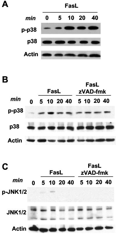
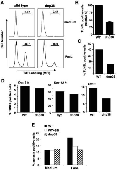
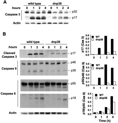
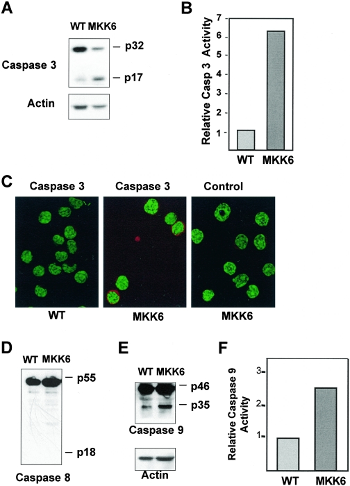
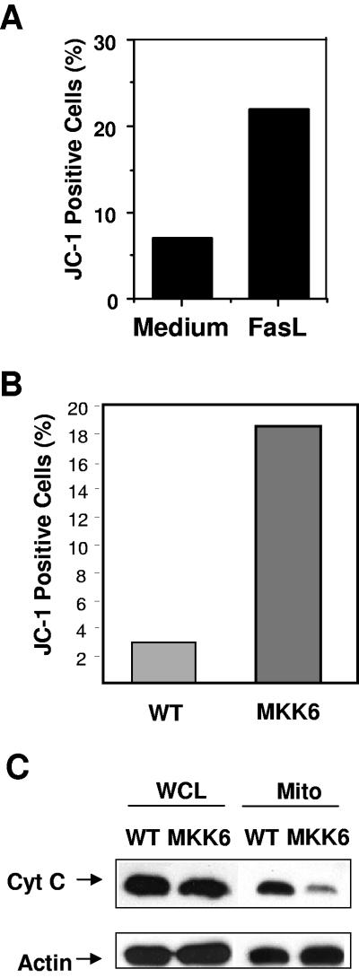
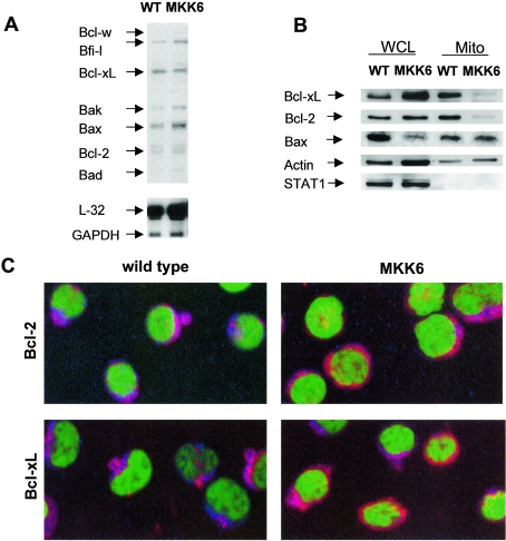
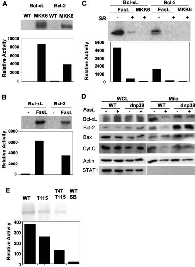
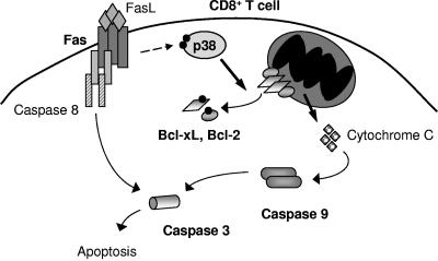
The p38 mitogen-activated protein kinase (MAPK) signaling pathway can be activated by a variety of stress stimuli such as UV radiation and osmotic stress. The regulation and role of this pathway in death receptor-induced apoptosis remain unclear and may depend on the specific death receptor and cell type. Here we show that binding of Fas ligand to Fas activates p38 MAPK in CD8+ T cells and that activation of this pathway is required for Fas-mediated CD8+ T-cell death. Active p38 MAPK phosphorylates Bcl-xL and Bcl-2 and prevents the accumulation of these antiapoptotic molecules within the mitochondria. Consequently, a loss of mitochondrial membrane potential and the release of cytochrome c lead to the activation of caspase 9 and, subsequently, caspase 3. Therefore, the activation of p38 MAPK is a critical link between Fas and the mitochondrial death pathway and is required for the Fas-induced apoptosis of CD8+ T cells.


Figures

FIG. 1.
FIG. 1.
Activation of p38 MAPK in response to FasL in CD8+ T cells. (A) Wild-type CD8+ T cells were cultured for 30 min in medium alone, prior to the addition of FasL (200 ng/ml) for the times indicated. Cells were then lysed, and a Western blot was performed for activated p38 MAPK (phospho-p38 [p-p38]), total p38 MAPK, and actin (as a loading control). (B) Wild-type CD8+ T cells were cultured in the presence or absence of zVAD-fmk (50 μM) for 30 min, prior to the addition of FasL (200 ng/ml) for the times indicated. Phospho-p38 and total p38 were examined by Western blot analysis as described for panel A. (C) Wild-type CD8+ T cells were cultured as described for panel B. Phospho-JNK1 and -JNK2 (p-JNK1/2) and total JNK were examined by Western blot analysis.
FIG. 2.
FIG. 2.
Activation of p38 MAPK is necessary for FasL-induced death in CD8+ T cells. (A) Freshly isolated CD8+ T cells from wild-type mice or dnp38 transgenic mice were cultured in medium alone or treated with FasL (200 ng/ml) for 4 h prior to detection of apoptosis via TUNEL and flow cytometry. The percentage of TdT-labeled cells is indicated. MFI, mean fluorescence intensity. This is a representative experiment, and panel B shows the mean percentage of TUNEL-positive (TdT labeled) cells (in response to FasL) relative to wild-type (WT) cells in four independent experiments (n = 4). Error bars represent standard error. (C) Wild-type and dnp38 CD8+ T cells were treated with a higher dose of FasL (400 ng/ml) for 4 h. Apoptosis was determined by TUNEL assay. (D) CD8+ T cells were treated with dexamethasone (Dex; 1 μM) for 3 or 12 h or with TNF-α (50 ng/ml) for 12 h. Apoptosis was determined by TUNEL assay. (E) CD8+ T cells from wild-type and dnp38 transgenic mice were treated with FasL (200 ng/ml). Wild-type CD8+ T cells were also treated with FasL (200 ng/ml) in the presence of the p38 MAPK inhibitor SB203580 (5 μM) (WT+SB). Apoptosis was examined after 2 h by Annexin V staining and flow cytometry.
FIG. 3.
FIG. 3.
Inhibition of p38 MAPK in CD8+ T cells prevents activation of caspase 3 and caspase 9 in response to FasL. (A) CD8+ T cells from wild-type or dnp38 transgenic mice were treated with FasL for the indicated time periods. Cells were then lysed, and a Western blot was performed using an antibody that recognizes both the full-length (p32) and cleaved (p17) caspase 3. (B) CD8+ T cells from wild-type (WT) or dnp38 transgenic mice were treated with FasL for the indicated time periods and lysed, and whole extracts were used for Western blots using antibodies against cleaved caspase 3 (p17), full-length (p46) and cleaved (p35) caspase 9, or full-length (p55) and cleaved (p18) caspase 8. Actin was examined as a loading control. Densitometry analysis was performed, and relative values for caspase 3 and for the cleaved/full-length-form ratio of caspase 8 and caspase 9 are shown. Results shown are representative of four independent experiments.
FIG. 4.
FIG. 4.
Activation of p38 MAPK alone is sufficient to induce activation of caspase 3 and caspase 9, but not caspase 8, in CD8+ T cells. (A) Whole-cell lysates of freshly isolated CD8+ T cells from wild-type (WT) or MKK6(Glu) transgenic mice were analyzed by Western blotting for full-length (p32) and cleaved (p17) caspase 3. Actin was examined as a loading control. (B) Relative caspase 3 activity of CD8+ T cells from wild-type or MKK6(Glu) transgenic mice as measured by cleavage of a luminescent caspase 3 substrate. (C) Freshly isolated CD8+ T cells from wild-type and MMK6(Glu) transgenic mice were used to examined active caspase 3 by immunostaining and confocal microscopy analysis using an anti-cleaved caspase 3 (p17) antibody and a secondary antibody (red). YOYO (green) was used as a nuclear marker. Staining with the secondary antibody (Control) in MKK6(Glu) CD8+ T cells is also shown. (D and E) Whole-cell lysates of CD8+ T cells from wild-type or MKK6(Glu) transgenic mice were analyzed by Western blotting for full-length (p55) and cleaved (p18) caspase 8 (D) or full-length (p46) and cleaved (p35) caspase 9 (E). Actin was examined as a loading control. (F) Relative caspase 9 activity of CD8+ T cells from wild-type or MKK6(Glu) transgenic mice as measured by cleavage of a luminescent caspase 9 substrate. Results are representative of three independent experiments.
FIG. 5.
FIG. 5.
CD8+ T cells from MKK6(Glu) transgenic mice have decreased mitochondrial membrane potential and decreased mitochondrial cytochrome c. (A) Wild-type CD8+ T cells were incubated with medium alone or FasL (200 ng/ml). After 4 h, cells were stained with JC-1 dye and analyzed by flow cytometry. The percentages of cells containing JC-1 green fluorescence (indicating a loss of mitochondrial membrane potential) are shown. (B) Freshly isolated CD8+ T cells from wild-type (WT) or MKK6(Glu) transgenic mice were stained with JC-1 dye and analyzed by flow cytometry. (C) Whole-cell lysates (WCL) or mitochondrial lysates (Mito) from wild-type or MKK6(Glu) CD8+ T cells were analyzed by Western blotting for cytochrome c (Cyt C) or actin (as a loading control). The results shown are representative of three independent experiments.
FIG. 6.
FIG. 6.
Activation of p38 MAPK in CD8+ T cells prevents mitochondrial accumulation of Bcl-2 and Bcl-xL. (A) Total RNA from freshly purified wild-type (WT) or MKK6(Glu) CD8+ T cells was analyzed for expression of various Bcl-2 family members by RPA using the mAPO-2 template kit (BD PharMingen). L-32 and GAPDH were examined as controls for loading. (B) Whole-cell lysates (WCL) or mitochondria lysates (Mito) from wild-type or MKK6(Glu) CD8+ T cells were analyzed by Western blotting for Bcl-xL, Bcl-2, and Bax. We also examined the levels of actin (as a loading control) and STAT1 (as a marker of cytoplasmic contamination). (C) Cytospun CD8+ T cells from wild-type or MKK6(Glu) transgenic mice were stained with antibodies against either Bcl-2 or Bcl-XL (red), YOYO nuclear dye (green), and MitoTracker mitochondrial dye (blue). Colocalization of Bcl-2 or Bcl-xL with MitoTracker is displayed as a pink/purple color, while cytosolic Bcl-2 and Bcl-xL appear red. Cells were visualized by confocal microscopy at a ×60 magnification. The results shown are representative of at least two independent experiments.
FIG. 7.
FIG. 7.
p38 MAPK from MKK6(Glu) transgenic or FasL-treated wild-type, CD8+ T cells can phosphorylate Bcl-2 and Bcl-xL. (A) Total p38 MAPK was immunoprecipitated from whole-cell lysates of wild-type (WT) or MKK6(Glu) CD8+ T cells and incubated with recombinant Bcl-xL or Bcl-2 substrates in vitro in the presence of [γ-32P]ATP. Substrate phosphorylation following SDS-PAGE was detected by autoradiography (upper panel) and quantitated by PhosphorImager (lower panel). (B) Wild-type CD8+ T cells were cultured in the presence (FasL) or absence (−) of FasL for 2 h prior to lysis and immunoprecipitation of total p38 MAPK. The ability of p38 MAPK to phosphorylate Bcl-xL and Bcl-2 was analyzed as described for panel A. (C) Total p38 MAPK was immunoprecipitated from wild-type CD8+ T cells treated for 2 h with FasL (FasL) or freshly isolated MKK6(Glu) CD8+ T cells (MKK6). The phosphorylation of Bcl-xL and Bcl-2 in vitro was analyzed as described for panel A, in the presence (+) or absence (−) of the p38 MAPK inhibitor SB203580 (2.5 μM). (D) CD8+ T cells from wild-type or dnp38 transgenic mice were treated with FasL (+) or medium alone (−) for 2 h. Whole-cell lysates (WCL) or mitochondrial lysates (Mito) were prepared and examined for the presence of Bcl-xL, Bcl-2, Bax, and cytochrome c (Cyt C) by Western blot analysis. We also analyzed the levels of actin (as a loading control) and STAT1 (as a marker of cytoplasmic contamination). (E) Total p38 MAPK was immunoprecipitated from freshly isolated MKK6(Glu) CD8+ T cells (MKK6). The phosphorylation of wild-type Bcl-xL, the Thr115Ala Bcl-xL mutant (Thr115) and the Thr115Ala Thr47Ala double Bcl-xL mutant (T47T115) by p38 MAPK in vitro was analyzed as described for panel A. Phosphorylation of wild-type Bcl-xL in the presence of SB203580 (WT/SB) is also shown. The results shown are representative of at least two independent experiments.
FIG. 8.
FIG. 8.
Role for Fas-induced p38 MAPK activity in apoptosis of CD8+ T cells.

References

    1. Alvarado-Kristensson, M., F. Melander, K. Leandersson, L. Ronnstrand, C. Wernstedt, and T. Andersson. 2004. p38-MAPK signals survival by phosphorylation of caspase-8 and caspase-3 in human neutrophils. J. Exp. Med. 199:449-458. - PMC - PubMed
    1. Braz, J. C., O. F. Bueno, Q. Liang, B. J. Wilkins, Y.-S. Dai, S. Parsons, J. Braunwart, B. J. Glascock, R. Klevitsky, T. F. Kimball, T. E. Hewett, and J. D. Molkentin. 2003. Targeted inhibition of p38 MAPK promotes hypertrophic cardiomyopathy through upregulation of calcineurin-NFAT signaling. J. Clin. Investig. 111:1475-1486. - PMC - PubMed
    1. Brenner, B., U. Koppenhoefer, C. Weinstock, O. Linderkamp, F. Lang, and E. Gulbins. 1997. Fas- or ceramide-induced apoptosis is mediated by a Rac1-regulated activation of Jun N-terminal kinase/p38 kinases and GADD153. J. Biol. Chem. 272:22173-22181. - PubMed
    1. Cahill, M. A., M. E. Peter, F. C. Kischkel, A. M. Chinnaiyan, V. M. Dixit, P. H. Krammer, and A. Nordheim. 1996. CD95 (APO-1/Fas) induces activation of SAP kinases downstream of ICE-like proteases. Oncogene 13:2087-2096. - PubMed
    1. Cohen, P. L., and R. A. Eisenberg. 1991. Lpr and gld: single gene models of systemic autoimmunity and lymphoproliferative disease. Annu. Rev. Immunol. 9:243-269. - PubMed

Publication types

MeSH terms