. 2006 Mar;26(6):2273-85.
doi: 10.1128/MCB.26.6.2273-2285.2006.

CARM1 regulates proliferation of PC12 cells by methylating HuD

Affiliations

CARM1 regulates proliferation of PC12 cells by methylating HuD

Tatsuji Fujiwara et al. Mol Cell Biol. 2006 Mar.

Abstract

HuD is an RNA-binding protein that has been shown to induce neuronal differentiation by stabilizing labile mRNAs carrying AU-rich instability elements. Here, we show a novel mechanism of arginine methylation of HuD by coactivator-associated arginine methyltransferase 1 (CARM1) that affected mRNA turnover of p21cip1/waf1 mRNA in PC12 cells. CARM1 specifically methylated HuD in vitro and in vivo and colocalized with HuD in the cytoplasm. Inhibition of HuD methylation by CARM1 knockdown elongated the p21cip1/waf1 mRNA half-life and resulted in a slow growth rate and robust neuritogenesis in response to nerve growth factor (NGF). Methylation-resistant HuD bound more p21cip1/waf1 mRNA than did the wild type, and its overexpression upregulated p21cip1/waf1 protein expression. These results suggested that CARM1-methylated HuD maintains PC12 cells in the proliferative state by committing p21cip1/waf1 mRNA to its decay system. Since the methylated population of HuD was reduced in NGF-treated PC12 cells, downregulation of HuD methylation is a possible pathway through which NGF induces differentiation of PC12 cells.


Figures
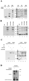
FIG. 1.
FIG. 1.
CARM1-methylated Arg236 of HuD in vitro. (A) Recombinant histones H3 and H4 and GST-tagged HuDwt were methylated by either His6-PRMT1 or His6-CARM1. GST-HuD, histone H3, or histone H4 (3 μg each) was incubated with 1 μg of His-PRMTs in the presence of 1μCi of [3H]AdoMet. As reported, histone H4 was methylated chiefly by His6-PRMT1 (left panel, lanes 1 and 2) and histone H3 predominantly by His6-CARM1 (left panel, lanes 3 and 4). GST-HuDwt was a specific substrate for His6-CARM1, not His6-PRMT1 (left panel, lanes 5 and 6). The methylated band indicated by an arrow was a histone H3 dimer. The right panel shows Coomassie staining of the gel identical to that shown in the left panel. Each of the substrates (asterisks) and enzymes (arrowheads) are indicated. Lane M is a molecular size marker (in kilodaltons). (B) In vitro methylation assay of GST-HuD mutants. GST-HuD, -R236K, -R238K, and GST alone were incubated with CARM1. 3H-labeled bands were found in GST-HuDwt and GST-R238K to similar extents (left panel, lanes 2 and 4, arrowheads), while GST-R236K harboring a mutation at R236 (corresponding to R217 of HuR) was not 3H labeled by CARM1 (left panel, lane 3). The faster-migrating bands are presumed to be degradative products of the HuD portion. GST alone also failed to undergo methylation (left panel, lane 1). Input of the recombinant proteins was verified by Coomassie staining of the gel shown in the left panel (right panel). GST and GST-tagged proteins are indicated by asterisks and CARM1 by the arrow. (C) CARM1-methylated GST-HuD was blotted with anti-M/DMA. GST-HuD and GST-R236K were incubated with recombinant CARM1 (left panel, lanes 3 and 4) and PRMT1 (left panel, lane 5). Input of GST-HuD proteins was verified by Coomassie staining (right panel). (D) HuD harboring the R236K mutation was methylation defective. Cell extracts from HA-HuD- and HA-R236K-expressing PC12 cells were immunoprecipitated with anti-HA antibody, and the precipitated proteins were analyzed by immunoblotting with anti-M/DMA antibody (upper panel) or anti-HA antibody (lower panel). Only HA-HuD was detectable with anti-HA antibody, while HA-R236K was not, as expected.
FIG. 2.
FIG. 2.
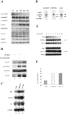
Arg236 of HuD was methylated by CARM1 in PC12 cells. (A) CARM1 expression was abrogated by stably expressing CARM1 shRNA in PC12 cells, as shown by immunocytochemistry using antiserum against CARM1 (green), phalloidin-actin (red), and DAPI (blue). The empty vector-transfected cells did show a dense signal in the nuclei and were moderately stained in the cytoplasm (a), while shRNA specific for CARM1 sequence significantly suppressed the nuclear staining (c). (B) Specific reduction of CARM1 level in one of the clones stably expressing CARM1 shRNA, as shown by immunoblot analysis. Total proteins isolated from the CARM1 shRNA-expressing PC12 cells and the parental cells were blotted with antibodies against CARM1, PRMT1, PRMT3, and actin. CARM1 shRNA did not affect PRMT1 and PRMT3 protein levels (right column). (C) Endogenous HuD was arginine methylated in the native PC12 cells but not in the CARM1 cells. Whole-cell extracts from parental and CARM1 PC12 cells were loaded directly (lower panel) or immunoprecipitated with either anti-M/DMA antibody or control IgG (upper panel). These samples were analyzed by immunoblotting with anti-HuD antibody. Anti-M/DMA antibody precipitated endogenous HuD from mock-transfected PC12 cell lysate (lane 2) but not from the lysate of CARM1 cell line 15 (lane 3). The fraction precipitated with control IgG1 contained no HuD immunoreactivity (lane 1). CTR, control empty vector-expressing cells; siRNA, CARM1 shRNA-expressing cells.
FIG. 3.
FIG. 3.
Subcellular localization of CARM1 in PC12 cells. (A) Subcellular localization of CARM1 in PC12 cells was confirmed by HRP-DAB detection. In HeLa cells, CARM1 immunoreactivities were found to occur exclusively in the nuclei, but CARM1 exhibited cytoplasmic localization, as well as intense nuclear localization, in PC12 cells. When antiserum adsorbed with recombinant CARM1 was applied to the PC12 cells, both cytoplasmic and nuclear immunoreactivities were lost. The lower panels show high-power views of the cells shown in the upper panels. (B) Fluorescence immunocytochemistry using anti-CARM1 (a and d) (green) and anti-HuD (b and e) (red) antibodies with mock-transfected PC12 cells (a and b) and CARM1 cells (d and e). Cytoplasmic colocalization of both immunoreactivities (yellow) was found with the mock-transfected cells (c), whereas no remarkable colocalization was found with the CARM1 cells (f). The colocalization represented the perinuclear pattern and declined at the cell peripheries. Some cells had colocalizing spots in the growing neurites (data not shown). siRNA, CARM1 shRNA-expressing cells. (C) Immunocytochemistry using goat anti-HuD antibody. The same distribution pattern as described for panel B was observed.
FIG. 4.
FIG. 4.
CARM1 depletion in PC12 cells exclusively induces p21cip1/waf1 expression. (A) Protein expression patterns of HuD-regulating genes in the wild-type (W), mock-transfected (M), and CARM1 PC12 cells, which were all cultured under the same growth condition. The whole-cell lysates of these cells were blotted with anti-CARM1, -PRMT1, -PRMT3, -p21cip1/waf1, -GAP43, -tau, -p27, and -actin antibodies. Note that p21cip1/waf1 protein levels significantly increased in CARM1 clones 15, 16, and 33. These cell lines also represented a slight increment of basal protein level of GAP43. GAPDH served to monitor equal loading and transfer of samples. (B) Effects of exogenous HA-HuD and HA-R236K on inducing p21cip1/waf1 expression. PC12 cells were transiently transfected with HA-HuD or HA-R236K, and total lysates were blotted with anti-p21cip1/waf1, -GAP43, and -actin antibodies. The p21cip1/waf1 protein level was significantly increased in HA-R236K-expressing cells (top panel, lane 3). The same levels of HA-tagged proteins were monitored by anti-HA blotting. (C) Upregulation of p21cip1/waf1 mRNA in CARM1 clone 15. Northern blot analyses were performed with total RNAs extracted from the parental, CARM1, and NGF-treated parental PC12 cells. The basal p21cip1/waf1 transcript level in CARM1 cells (middle column) was about threefold higher than that in the parental cells (left column), which was comparable to that in the native cells treated with NGF for 2 days (2d) (right column). Differences (n-fold) are indicated between the blots. GAP43 and tau transcripts made almost no difference between the parental and CARM1 cells or NGF-upregulated p21cip1/waf1, GAP43, and tau transcript levels (right column). GAPDH mRNA served to monitor equal loading and transfer of samples. (D) The basal transcription level of the p21cip1/waf1 gene was not affected by CARM1 depletion. Nuclei from parental, CARM1, and NGF-treated PC12 cells were isolated for nuclear run-on assay to monitor transcription rates for the genes indicated. The PCR product (1 μg) corresponding to each gene was blotted onto nitrocellulose filters. GAPDH was employed as a control gene (not induced by NGF), GAP43 as a positive control gene (induced by NGF), and green fluorescent protein (GFP) as a negative control. (E) p21cip1/waf1 mRNA was stabilized in CARM1 PC12 cells to the extent of NGF-treated native PC12 cells. The time at which actinomycin D was added to the media is indicated as 0 h. Total RNA was prepared at each indicated time and analyzed by Northern blot analysis. (F) Quantification of half-life of p21cip1/waf1 mRNA for each cell group. Densitometric values of p21cip1/waf1 at every time point were plotted on a logarithmic scale. The half-life of p21cip1/waf1 mRNA is formulated as the time at which the density was reduced to half that of the initial density. Data represent the means ± standard errors of the means from three independent experiments.
FIG. 5.
FIG. 5.
Unmethylated HuD exhibited a higher binding capacity for p21cip1/waf1 mRNA. (A) Detection by RT-PCR of p21 mRNA in materials that were precipitated with anti-HA antibody from HA-HuD- and HA-R236K-expressing cells. Control IgG1 did not yield the amplified product of p21cip1/waf1 (top panel, lane 1) from HA-HuD-expressing cells. HA-R236K precipitated with anti-HA antibody yielded 5.8 times more PCR product of p21cip1/waf1 than HA-HuD (top panel, lanes 2 and 3). Differences (n-fold) are indicated between the blots. NGF treatment propagated the amplified product of p21cip1/waf1 from HA-HuD to the extent of HA-R236K (top panel, lane 4). Background detection of GAPDH-amplified product from each of the precipitated materials served to monitor equal use of material in each IP (middle panel). (B) NGF downregulated the methylated population of HuD. Whole-cell extracts from the untreated PC12 cells and the cells treated with NGF for 3 days were loaded directly (lanes 3 and 4) or subjected to IP with anti-M/DMA antibody (lanes 1 and 2), and they were blotted with anti-HuD antibody. Antiactin antibody blotting of the total lysates confirmed the same protein load (lanes 5 and 6).
FIG. 6.
FIG. 6.
Effects of CARM1 loss on proliferation and NGF-induced neurite outgrowth. (A) Comparison of levels of proliferation in parent, mock-transfected, and CARM1-depleted cells. PC12 cells were plated on 6-well dishes at a density of 1 × 10⁵ cells per well. The cells were then cultured in growth medium and counted by a hematocytometer at every 24-h interval. Cell numbers represent the mean values from triplicate experiments. CARM1-defective cells (no. 15, 16, and 33) showed slower growth rates than parental and mock-transfected cells. (B) CARM1 depletion promoted a susceptibility to NGF-induced neurite outgrowth. Parental PC12 cells and CARM1 cells were plated on collagen-coated, 4-well-chamber slides at a density of 1 × 10⁴ cells per well. Microscopic images of the parent and CARM1 PC12 cells (no. 15 and 33) are shown. Cultures were fixed at the indicated times after the onset of NGF exposure and stained with tetramethyl rhodamine isothiocyanate-labeled phalloidin (red). (C) Quantification of the results shown in panel B. The cells that had at least one neurite with a length covering two cell bodies in diameter was counted as neurite bearing. The error bars represent standard deviations from triplicate results. Approximately 200 cells were counted for each sample.
FIG. 7.
FIG. 7.
Distribution of CARM1-expressing cells in the adult mouse brain. Immunofluorescent double staining of the VZ was achieved by use of the antibodies to CARM1 (green) (a) and BrdU (red) (b). In the adult mouse brain, CARM1-expressing cells were mainly observed in the lateral VZ and dentate gyrus. Some CARM1-expressing cells in the lateral ventricular wall are costained with BrdU (c). LV, lateral ventricle.

Similar articles

Cited by

References

    1. Abe, R., E. Sakashita, K. Yamamoto, and H. Sakamoto. 1996. Two different RNA binding activities for the AU-rich element and the poly(A) sequence of the mouse neuronal protein mHuC. Nucleic Acids Res. 24:4895-4901. - PMC - PubMed
    1. Afonina, E., R. Stauber, and G. N. Pavlakis. 1998. The human poly(A)-binding protein 1 shuttles between the nucleus and the cytoplasm. J. Biol. Chem. 273:13015-13021. - PubMed
    1. Akamatsu, W., H. Fujihara, T. Mitsuhashi, M. Yano, S. Shibata, Y. Hayakawa, H. J. Okano, S. Sakakibara, H. Takano, T. Takano, T. Takahashi, T. Noda, and H. Okano. 2005. The RNA-binding protein HuD regulates neuronal cell identity and maturation. Proc. Natl. Acad. Sci. USA 102:4625-4630. - PMC - PubMed
    1. An, W., J. Kim, and R. G. Roeder. 2004. Ordered cooperative functions of PRMT1, p300, and CARM1 in transcriptional activation by p53. Cell 117:735-748. - PubMed
    1. Anderson, K. D., J. Sengupta, M. Morin, R. L. Neve, C. F. Valenzuela, and N. I. Perrone-Bizzozero. 2001. Overexpression of HuD accelerates neurite outgrowth and increases GAP-43 mRNA expression in cortical neurons and retinoic acid-induced embryonic stem cells in vitro. Exp. Neurol. 168:250-258. - PubMed

MeSH terms