. 2006 Mar;26(6):2408-18.
doi: 10.1128/MCB.26.6.2408-2418.2006.

Posttranslational regulation of tristetraprolin subcellular localization and protein stability by p38 mitogen-activated protein kinase and extracellular signal-regulated kinase pathways

Affiliations

Posttranslational regulation of tristetraprolin subcellular localization and protein stability by p38 mitogen-activated protein kinase and extracellular signal-regulated kinase pathways

Matthew Brook et al. Mol Cell Biol. 2006 Mar.

Abstract

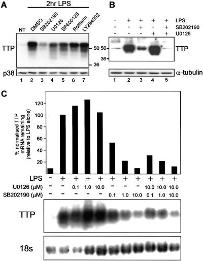
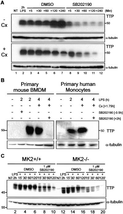
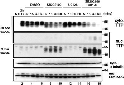
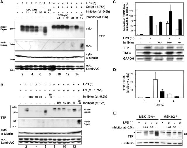
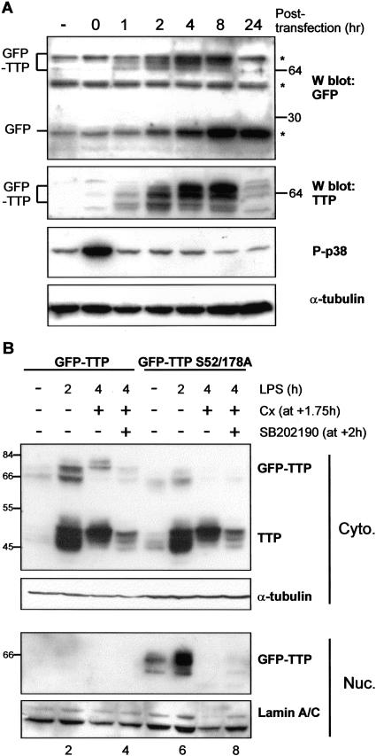
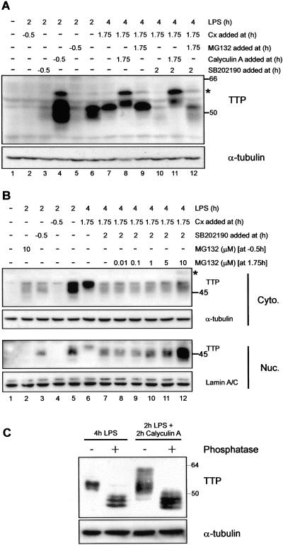
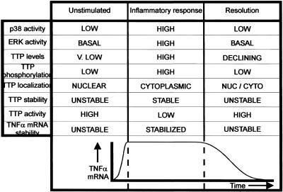
The p38 mitogen-activated protein kinase (MAPK) signaling pathway, acting through the downstream kinase MK2, regulates the stability of many proinflammatory mRNAs that contain adenosine/uridine-rich elements (AREs). It is thought to do this by modulating the expression or activity of ARE-binding proteins that regulate mRNA turnover. MK2 phosphorylates the ARE-binding and mRNA-destabilizing protein tristetraprolin (TTP) at serines 52 and 178. Here we show that the p38 MAPK pathway regulates the subcellular localization and stability of TTP protein. A p38 MAPK inhibitor causes rapid dephosphorylation of TTP, relocalization from the cytoplasm to the nucleus, and degradation by the 20S/26S proteasome. Hence, continuous activity of the p38 MAPK pathway is required to maintain the phosphorylation status, cytoplasmic localization, and stability of TTP protein. The regulation of both subcellular localization and protein stability is dependent on MK2 and on the integrity of serines 52 and 178. Furthermore, the extracellular signal-regulated kinase (ERK) pathway synergizes with the p38 MAPK pathway to regulate both stability and localization of TTP. This effect is independent of kinases that are known to be synergistically activated by ERK and p38 MAPK. We present a model for the actions of TTP and the p38 MAPK pathway during distinct phases of the inflammatory response.


Figures

FIG. 1.
FIG. 1.
TTP protein, but not mRNA expression, is synergistically regulated by the p38 MAPK and ERK pathways. (A and B) RAW 264.7 cells were either left unstimulated or stimulated with LPS for 2 h in the presence of either 0.1% DMSO, 1 μM SB202190, 10 μM U0126, 50 μM SP600125, 10 μM Rottlerin, or 10 μM LY294002. Whole-cell extracts were prepared, and TTP and tubulin were detected by Western blotting. Positions of molecular mass markers (in kilodaltons) are indicated. (C) RAW 264.7 cells were left unstimulated or stimulated with LPS for 2 h in the presence of 0.1% DMSO or the indicated concentrations of SB202190 or U0126. TTP mRNA was detected by Northern blotting and normalized against 18S rRNA. NT, untreated.
FIG. 2.
FIG. 2.
TTP protein stability is regulated by the p38 MAPK pathway. (A) RAW 264.7 cells were either left unstimulated, stimulated with LPS for 2 h, or stimulated with LPS for 2 h and then treated with 0.1% DMSO or 1 μM SB202190 for the times indicated (upper panel) or stimulated with LPS for 1.75 h and then treated with Cx (5 μg/ml) for 15 min prior to the addition of 0.1% DMSO or 1 μM SB202190 (lower panel) for the times indicated. (B) Primary mouse bone marrow-derived macrophages (BMDM) (left panel) or primary human monocytes (right panel) were either left unstimulated, stimulated with LPS for 2 h in the presence or absence of 1 μM SB202190, or stimulated with LPS for 1.75 h and then treated with Cx (5 μg/ml) for 15 min prior to the addition of 0.1% DMSO or 1 μM SB202190 for a further 2 h. (C) Immortalized wild-type or MK2−/− macrophages were either left unstimulated or stimulated with LPS for 1.75 h and then treated with Cx (5 μg/ml) for 15 min prior to the addition of 0.1% DMSO or 1 μM SB202190 for the times indicated; 100 μg wild type and 300 μg MK2−/− extract were loaded for blotting. In each case (A to C), whole-cell extracts were prepared and TTP and tubulin were detected by Western blotting. Positions of molecular mass markers (in kilodaltons) are indicated. NT, untreated.
FIG. 3.
FIG. 3.
TTP protein stability and subcellular localization are synergistically regulated by the p38 MAPK and ERK pathways. RAW 264.7 cells were either left unstimulated, stimulated with LPS for 2 h, or stimulated with LPS for 1.75 h and then treated with Cx (5 μg/ml) for 15 min prior to the addition of 0.1% DMSO, 1 μM SB202190, and/or 10 μM U0126 for the times indicated. Cytoplasmic (cyto.) and nuclear (nuc.) extracts were prepared, and TTP, tubulin, and lamin A/C were detected by Western blotting (tubulin was not detected in the nuclear fraction, and lamin A/C was not detected in the cytoplasmic fraction). Positions of molecular mass markers (in kilodaltons) are indicated. Equivalent exposures (expos.) (30 s) of the cytoplasmic and nuclear TTP signals and an extended exposure (3 min) of the nuclear TTP signal are shown. NT, untreated.
FIG. 4.
FIG. 4.
MSK-1 and -2 but not MNK-1 and -2 regulate TTP protein induction but not its stability or subcellular localization. (A) RAW 264.7 cells were either left unstimulated, stimulated with LPS for 2 h in the presence or absence of the indicated doses of CPG57380 (CPG), 1 μM SB202190 (SB), and/or 10 μM U0126 (U), or stimulated with LPS for 1.75 h and then treated with Cx (5 μg/ml) for 15 min prior to the addition of 0.1% DMSO, the indicated doses of CPG57380, 1 μM SB202190, and/or 10 μM U0126 for the times indicated. An extended exposure (expos.) (5 min) of the nuclear TTP signal is shown. (B) As for panel A, except that the inhibitors used were as follows: 25 μM H89, 5 μM Ro 31-8220, 1 μM SB202190, and/or 10 μM U0126. An extended exposure (5 min) of the nuclear TTP signal is shown. In each case (A and B), cytoplasmic (cyto.) and nuclear (nuc.) extracts were prepared, and TTP, tubulin, and lamin A/C were detected by Western blotting (tubulin was not detected in the nuclear fractions, and lamin A/C was not detected in the cytoplasmic fraction). (C) RAW 264.7 cells were either left unstimulated or treated with 0.1% DMSO, 10 μM MG132 (MG), 25 μM H89, or 5 μM Ro 31-8220 (Ro) for 30 min prior to being treated with LPS for 2 h. Total RNA was prepared, and 4 μg RNA/point was analyzed by RNase protection assay for the expression of TTP mRNA (black bars) and TNF-α mRNA (gray bars). (D) Wild-type (open bars) and MSK-1−/− MSK-2−/− macrophages (black bars) were either left unstimulated or treated with LPS for the times indicated. Total RNA was prepared, and TTP mRNA levels were measured using quantitative reverse transcription-PCR. Means ± standard deviations are shown for duplicate PCRs on two independent macrophage preparations per genotype. (E) Wild-type and MSK-1−/− MSK-2−/− macrophages were either left unstimulated or were treated with 0.1% DMSO, 25 μM H89, or 1 μM SB202190 for 30 min prior to being treated with LPS for 2 h. Whole-cell extracts were prepared for Western blotting. Positions of molecular mass markers (in kilodaltons) are indicated in each case. An asterisk indicates a nonspecific band.
FIG. 5.
FIG. 5.
TTP stability and subcellular localization are regulated by MK2-mediated phosphorylation of serines 52 and 178. (A) RAW 264.7 cells were either left untreated or transfected with peGFPc1-TTP and cultured for the times indicated following transfection. Whole-cell extracts were prepared, and GFP-TTP, GFP, phospho-p38 (P-p38) MAPK, and tubulin were detected by Western blotting (W blot). Asterisks indicate nonspecific bands detected by the anti-GFP antibody. (B) RAW 264.7 cells were transfected with either peGFPc1-TTP or peGFPc1-TTP-S52/178A and then cultured for 2 h. Cells were then either harvested, treated with LPS for 2 h, or treated with LPS for 1.75 h and then treated with 5 μg/ml Cx for 15 min prior to the addition of 0.1% DMSO or 1 μM SB202190 for a further 2 h. Cytoplasmic (cyto.) and nuclear (nuc.) extracts were prepared, and TTP, tubulin, and lamin A/C were detected by Western blotting (tubulin was not detected in the nuclear fractions, and lamin A/C was not detected in the cytoplasmic fraction). Positions of molecular mass markers (in kilodaltons) are indicated.
FIG. 6.
FIG. 6.
TTP turnover proceeds via the proteasome and is negatively regulated by p38 MAPK-mediated phosphorylation. (A) RAW 264.7 cells were either left unstimulated, treated with LPS for 2 h in the presence or absence of the indicated inhibitors, or treated with LPS for 1.75 h and then treated with 5 μg/ml Cx and/or 10 μM MG132 or 100 nM calyculin A for 15 min prior to the addition of 0.1% DMSO or 1 μM SB202190 for a further 2 h. Whole-cell extracts were prepared and TTP and tubulin were detected by Western blotting. Positions of molecular mass markers (in kilodaltons) are indicated. (B) RAW 264.7 cells were either left unstimulated, treated with LPS for 2 h in the presence or absence of 5 μg/ml Cx, 1 μM SB202190, or 10 μM MG132, or treated with LPS for 1.75 h and then treated with 5 μg/ml Cx and increasing concentrations of MG132 for 15 min prior to the addition of 1 μM SB202190 for a further 2 h. Cytoplasmic (Cyto.) and nuclear (Nuc.) extracts were prepared, and TTP, tubulin, and lamin A/C were detected by Western blotting (tubulin was not detected in the nuclear fraction, and lamin A/C was not detected in the cytoplasmic fraction). (C) RAW 264.7 cells were either LPS stimulated for 4 h or treated with LPS for 2 h and then treated with 10 nM calyculin A for a further 2 h. Complete cell extracts were prepared and either left untreated or incubated with 50 U shrimp alkaline phosphatase for 2 h at 37°C prior to Western blotting. Positions of molecular mass markers (in kilodaltons) are indicated. An asterisk indicates a novel ∼63-kDa form of TTP.
FIG. 7.
FIG. 7.
Model for the regulation of inflammation by p38 MAPK and TTP. NUC, nuclear; CYTO, cytoplasmic; V, very.

References

    1. Bakheet, T., B. R. Williams, and K. S. Khabar. 2003. ARED 2.0: an update of AU-rich element mRNA database. Nucleic Acids Res. 31:421-423. - PMC - PubMed
    1. Blackshear, P. J., W. S. Lai, E. A. Kennington, G. Brewer, G. M. Wilson, X. Guan, and P. Zhou. 2003. Characteristics of the interaction of a synthetic human tristetraprolin tandem zinc finger peptide with AU-rich element-containing RNA substrates. J. Biol. Chem. 278:19947-19955. - PubMed
    1. Brewer, B. Y., J. Malicka, P. J. Blackshear, and G. M. Wilson. 2004. RNA sequence elements required for high affinity binding by the zinc finger domain of tristetraprolin: conformational changes coupled to the bipartite nature of Au-rich MRNA-destabilizing motifs. J. Biol. Chem. 279:27870-27877. - PubMed
    1. Bridges, D., and G. B. Moorhead. 2005. 14-3-3 proteins: a number of functions for a numbered protein. Sci. STKE 2005:re10. - PubMed
    1. Brook, M., G. Sully, A. R. Clark, and J. Saklatvala. 2000. Regulation of tumour necrosis factor alpha mRNA stability by the mitogen-activated protein kinase p38 signalling cascade. FEBS Lett. 483:57-61. - PubMed

Publication types

MeSH terms