. 2005 Jan 1;61(Pt 1):75-8.
doi: 10.1107/S1744309104027241. Epub 2004 Dec 2.

Crystallization and preliminary X-ray crystallographic analysis of agkicetin-C from Deinagkistrodon acutus venom

Affiliations

Crystallization and preliminary X-ray crystallographic analysis of agkicetin-C from Deinagkistrodon acutus venom

Gufeng Xu et al. Acta Crystallogr Sect F Struct Biol Cryst Commun. .

Abstract

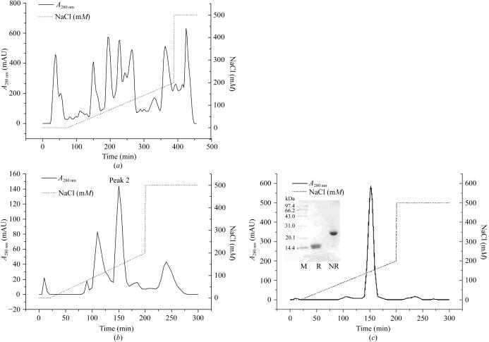
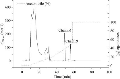
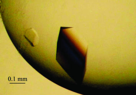
The crystallization and preliminary crystallographic analysis of agkicetin-C, a well known platelet glycoprotein Ib (GPIb) antagonist from the venom of Deinagkistrodon acutus found in Anhui Province, China is reported. Crystals of agkicetin-C suitable for structure determination were obtained from 1.8 M ammonium sulfate, 40 mM MES pH 6.5 with 2%(v/v) PEG 400. Interestingly, low buffer concentrations of MES seem to be necessary for crystal growth. The crystals of agkicetin-C belong to space group C2, with unit-cell parameters a = 177.5, b = 97.7, c = 106.8 A, beta = 118.5 degrees, and diffract to 2.4 A resolution. Solution of the phase problem by the molecular-replacement method shows that there are four agkicetin-C molecules in the asymmetric unit, with a VM value of 3.4 A3 Da(-1), which corresponds to a high solvent content of approximately 64%. Self-rotation function calculations show a single well defined non-crystallographic twofold axis with features that may represent additional elements of non-crystallographic symmetry.


Figures

Figure 1
Figure 1
Purification of agkicetin-C. (a) 1 g crude venom from D. acutus was dissolved in 20 ml buffer A (20 mM Tris–HCl pH 8.0) and centrifuged at 12 000 rev min−1 for 15 min. The supernatant was applied onto a DEAE-Sepharose Fast Flow column (1.6 × 40 cm) pre-equilibrated with buffer A. The column was washed at a flow rate of 120 ml h−1 initially with buffer A until peaks 1 and 2 were eluted and then with an 800 ml linear NaCl gradient (0–200 mM in buffer A). Finally, the column was exhaustively eluted with a high concentration of NaCl (500 mM in buffer A). Peak 4 containing agkicetin-C was pooled. (b) Peak 4 mentioned above was condensed and dialyzed against buffer B (50 mM sodium citrate pH 5.0) overnight at 277 K and then applied onto a CM-Sepharose Fast Flow column (1.6 × 40 cm) pre-equilibrated with buffer B. A similar strategy to the linear NaCl gradient used above was applied with buffer B and peak 2 was pooled. (c) Peak 2 mentioned above was condensed and dialyzed against buffer A overnight at 277 K and then applied onto a Mono Q HR 5/5 column (3.6 × 100 mm) pre-equilibrated with buffer A. The column was washed at a flow rate of 60 ml h−1 initially with 5 ml buffer A and then with a linear NaCl gradient (0–200 mM in buffer A). Finally, the column was eluted with a high concentration of NaCl (500 mM in buffer A) as mentioned above. The main fraction collected proved to be agkicetin-C.
Figure 2
Figure 2
Preparation of reduced and S-pyridylethylated agkicetin-C. 3 mg agkicetin-C was dissolved in 0.5 ml 0.5 M Tris–HCl buffer pH 8.0 containing 6 M guanidine hydrochloride and 2 mM EDTA. 7 mg dithiothreitol was added to the protein solution, mixed and then incubated for 3 h at 323 K. After the addition of 4-vinylpyridine with a 3:1 molar ratio of 4-vinylpyridine:dithiothreitol, the mixture was further incubated for another 3 h at 323 K and then dialyzed against distilled water and 0.1% TFA solution. Finally, the mixture was applied onto a Delta-Pak C4 column (3.9 × 150 mm) and sequentially eluted at room temperature with two linear gradients of acetonitrile solution (0–20% and 20–50% containing 0.1% TFA).
Figure 3
Figure 3
Crystals of agkicetin-C from 1.8 M ammonium sulfate, 2% PEG 400, 40 mM MES pH 6.5.

Similar articles

Cited by

References

    1. Andrews, R. K. & Berndt, M. C. (2000). Toxicon, 38, 775–791. - PubMed
    1. Andrews, R. K., Kroll, M. H., Ward, C. M., Rose, J. W., Scarborough, R. M., Smith, A. I., Lopez, J. A. & Berndt, M. C. (1996). Biochemistry, 35, 12629–12639. - PubMed
    1. Atoda, H., Hyuga, M. & Morita, T. (1991). J. Biol. Chem.266, 14903–14911. - PubMed
    1. Bartels K. S. & Klein C. (2003). The automar Manual, version 1.4. Norderstedt, Germany: marresearch GmbH.
    1. Brünger, A. T., Adams, P. D., Clore, G. M., Delano, W. L., Gros, P., Grosse-Kunstleve, R. W., Kuszewski, J., Nilges, M., Pannu, N. S., Read, R. J., Rice, L. M., Simonson, T. & Warren, G. L. (1998). Acta Cryst. D54, 905–921. - PubMed

Publication types

MeSH terms