

Crystallization and preliminary X-ray crystallographic analysis of peptide deformylase (PDF) from Bacillus cereus in ligand-free and actinonin-bound forms
- PMID: 16508119
- PMCID: PMC1952381
- DOI: 10.1107/S1744309104032440
Crystallization and preliminary X-ray crystallographic analysis of peptide deformylase (PDF) from Bacillus cereus in ligand-free and actinonin-bound forms
Abstract
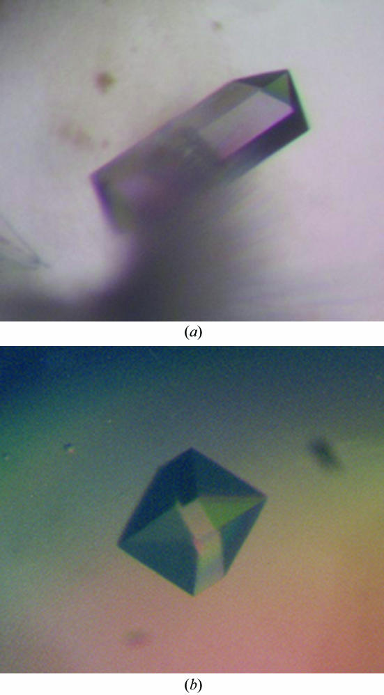
In bacteria, protein expression initiates with an N-formyl group and this needs to be removed in order to ensure proper bacterial growth. These formylation and deformylation processes are unique to eubacteria; therefore, inhibition of these would provide a novel antibacterial therapy. Deformylation is carried out by peptide deformylase (PDF). PDF from Bacillus cereus, one of the major pathogenic bacteria, was cloned into expression plasmid pET-28a (Novagen), overexpressed in Escherichia coli BL21 (DE3) and purified to high quality. Crystals have been obtained of both ligand-free PDF and PDF to which actinonin, a highly potent naturally occurring inhibitor, is bound. Both crystals belong to space group P2(1)2(1)2(1), with unit-cell parameters a = 42.72, b = 44.04, c = 85.19 A and a = 41.31, b = 44.56, c = 84.47 A, respectively. Diffraction data were collected to 1.7 A resolution for the inhibitor-free crystals and to 2.0 A resolution for the actinonin-bound crystals.
Figures
Similar articles
-
Co-crystallization of Leptospira interrogans peptide deformylase with a potent inhibitor and molecular-replacement schemes with eight subunits in an asymmetric unit.Acta Crystallogr D Biol Crystallogr. 2004 Jan;60(Pt 1):137-9. doi: 10.1107/s0907444903022480. Epub 2003 Dec 18. Acta Crystallogr D Biol Crystallogr. 2004. PMID: 14684909
-
Co-crystallization of Staphylococcus aureus peptide deformylase (PDF) with potent inhibitors.Acta Crystallogr D Biol Crystallogr. 2002 Dec;58(Pt 12):2153-6. doi: 10.1107/s090744490201569x. Epub 2002 Nov 23. Acta Crystallogr D Biol Crystallogr. 2002. PMID: 12454484
-
Crystallization and preliminary X-ray crystallographic analysis of peptide deformylase from Pseudomonas aeruginosa.Acta Crystallogr D Biol Crystallogr. 2002 Oct;58(Pt 10 Pt 2):1874-5. doi: 10.1107/s0907444902013835. Epub 2002 Sep 28. Acta Crystallogr D Biol Crystallogr. 2002. PMID: 12351843
-
Ligand-induced changes in the structure and dynamics of Escherichia coli peptide deformylase.Biochemistry. 2009 Aug 18;48(32):7595-607. doi: 10.1021/bi900600b. Biochemistry. 2009. PMID: 19627112 Free PMC article.
-
Bacterial Peptide deformylase inhibitors: a new class of antibacterial agents.Curr Med Chem. 2005;12(14):1607-21. doi: 10.2174/0929867054367194. Curr Med Chem. 2005. PMID: 16022661 Review.
References
-
- Adams, J. M. (1968). J. Mol. Biol.33, 571–589. - PubMed
-
- Groche, D., Becker, A., Schlichting, I., Kabsch, W., Schultz, S. & Wagner, A. F. V. (1998). Biochem. Biophys. Res. Commun.246, 342–346. - PubMed
-
- Guilloteau, J.-P., Mathieu, M., Giglione, C., Blanc, V., Dupuy, A., Chevrier, M., Gil, P., Famechon, A., Meinnel, T. & Mikol, V. (2002). J. Mol. Biol.320, 951–962. - PubMed
Publication types
MeSH terms
Substances
LinkOut - more resources
Full Text Sources
Other Literature Sources
