

In vitro organ culture of the bovine intervertebral disc: effects of vertebral endplate and potential for mechanobiology studies
- PMID: 16508544
- PMCID: PMC7187957
- DOI: 10.1097/01.brs.0000201302.59050.72
In vitro organ culture of the bovine intervertebral disc: effects of vertebral endplate and potential for mechanobiology studies
Abstract
Study design: Whole bovine coccygeal discs were cultured under static load, with or without vertebral endplates (VEPs), and assessed for cell viability, biochemical stability, biosynthetic activity, and biosynthetic responsiveness to changes in mechanical load.
Objectives: To assess the effects of VEPs on biochemical and cellular stability of disc cells during in vitro culture of large disc explants. To determine whether cultured discs could respond to mechanical perturbation.
Summary of background data: Previous methods for culturing the intervertebral disc have focused on rabbit and rat discs, but the small size of these discs limits the relevance of these culture systems to the human condition. Bovine coccygeal discs have similar dimensions to the human lumbar disc (i.e., similar size and nominal stresses), but long-term culture of these discs has not been reported.
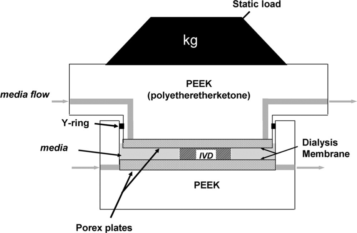
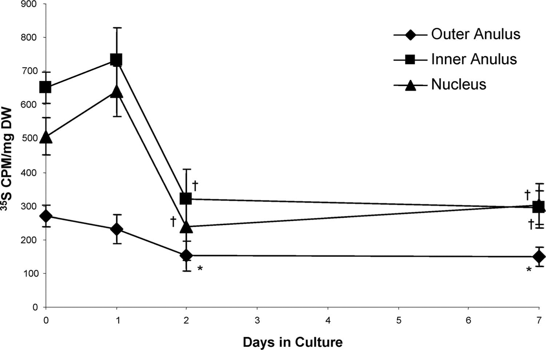
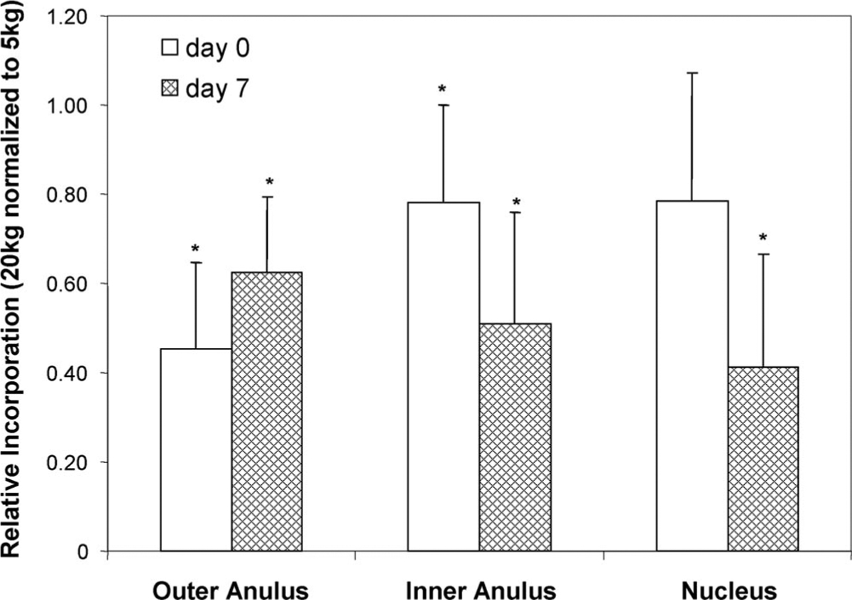
Methods: Bovine coccygeal discs were harvested with or without VEPs, cultured under static load (5 kg, approximately 0.25 MPa, in situ swelling pressure) for up to 1 week, and evaluated for changes in hydration, glycosaminoglycan content, cell viability, and biosynthetic activity. Additionally, the biochemical and biosynthetic response of discs cultured without VEP to increasing the load to a 20-kg (approximately 1 MPa, the estimated stress in human lumbar disc during heavy lifting) static load for 6 hours was assessed.
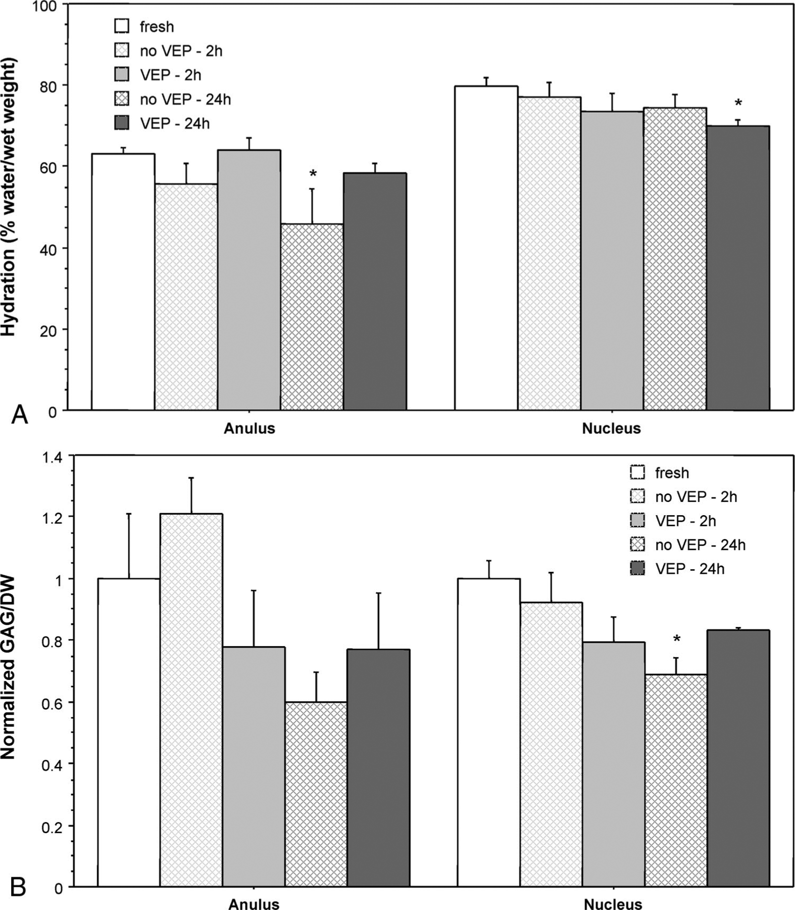
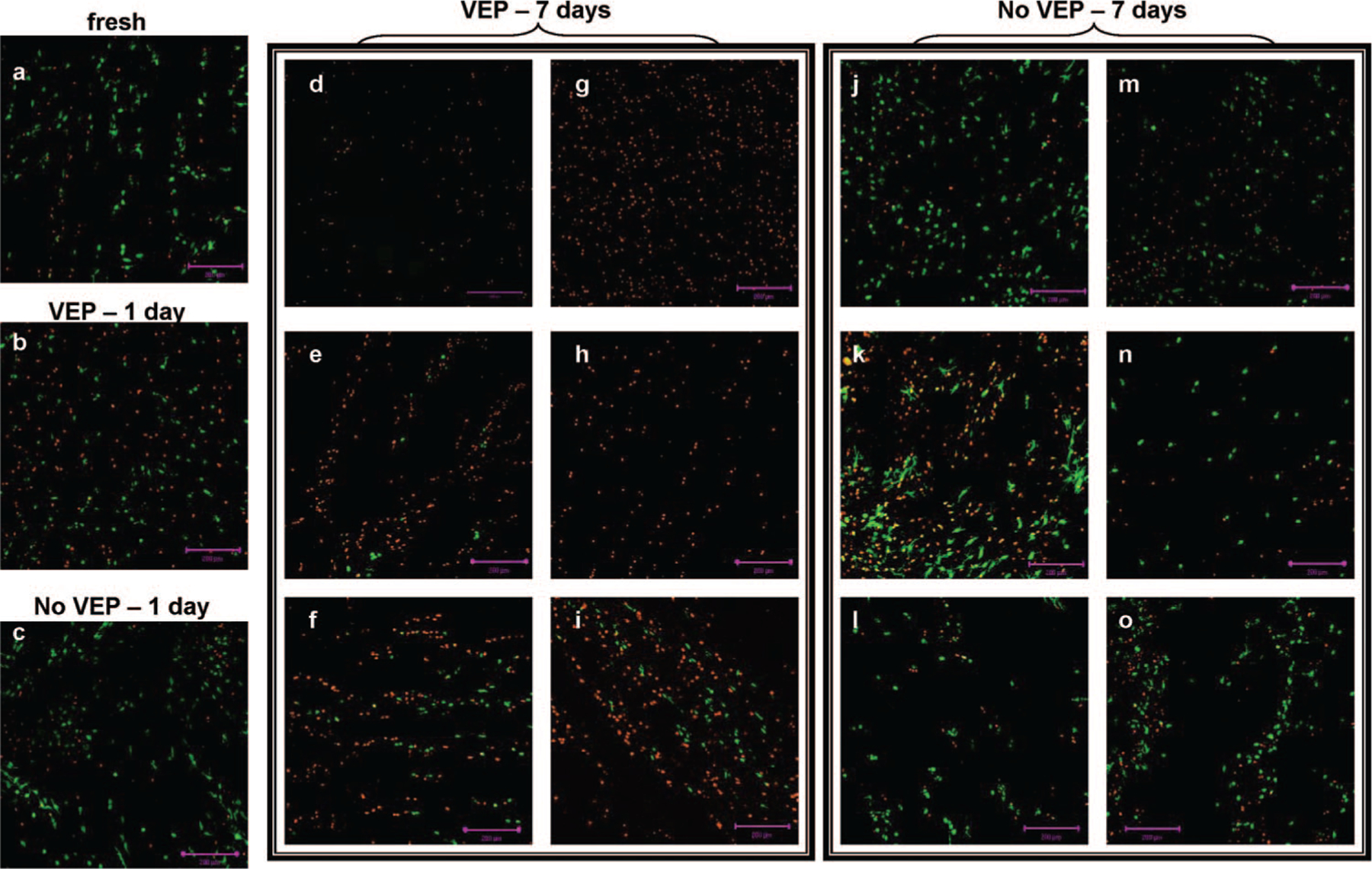
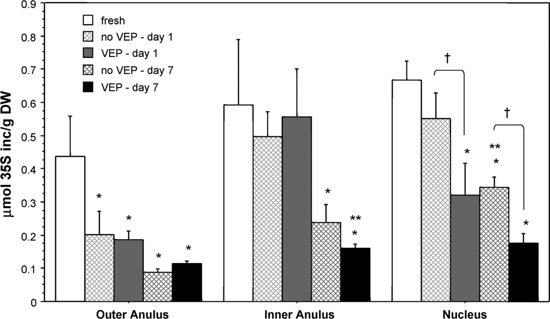
Results: During the first 24 hours, culturing discs with endplates was moderately better with regards to maintaining in situ anulus hydration and nucleus glycosaminoglycan levels. The endplates, however, obstructed media flow to the disc, resulting in a marked decrease in cell viability after 1 week of culture. Nucleus pulposus cell viability was maintained in discs cultured without endplates, but there was a significant drop in biosynthetic activity within 2 days of culture. Despite this drop, the disc cells in the discs without VEP remained biosynthetically responsive to changes in mechanical loading.
Conclusions: It is possible to maintain cell viability and the biosynthetic responsiveness of large discs for up to 1 week in vitro when the discs are cultured under static load and without VEP.
Figures
References
-
- Luo X, Pietrobon R, Sun SX, et al. Estimates and patterns of direct health care expenditures among individuals with back pain in the United States. Spine 2004;29:79–86. - PubMed
-
- Ala-Kokko L. Genetic risk factors for lumbar disc disease. Ann Med 2002; 34:42–7. - PubMed
-
- Battie MC, Videman T, Parent E. Lumbar disc degeneration: epidemiology and genetic influences. Spine 2004;29:2679–90. - PubMed
-
- Urban JP, Smith S, Fairbank JC. Nutrition of the intervertebral disc. Spine 2004;29:2700–9. - PubMed
Publication types
MeSH terms
Substances
Grants and funding
LinkOut - more resources
Full Text Sources
Research Materials
