

A distinct molecular profile associated with mucinous epithelial ovarian cancer
- PMID: 16508639
- PMCID: PMC2361366
- DOI: 10.1038/sj.bjc.6603003
A distinct molecular profile associated with mucinous epithelial ovarian cancer
Abstract
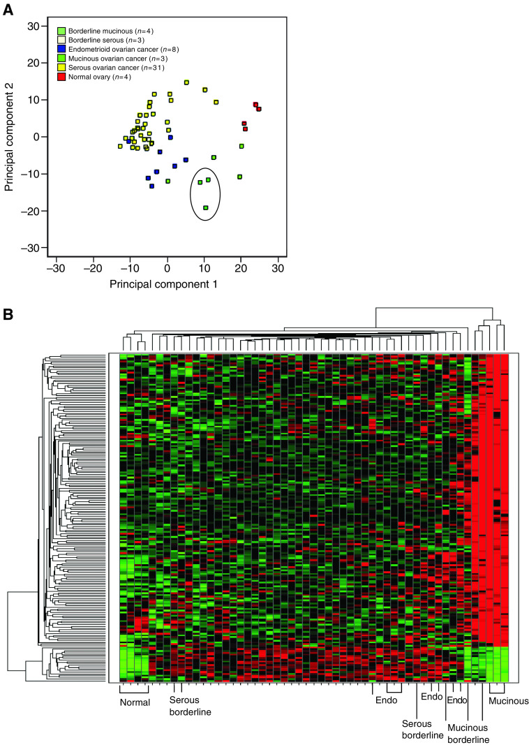
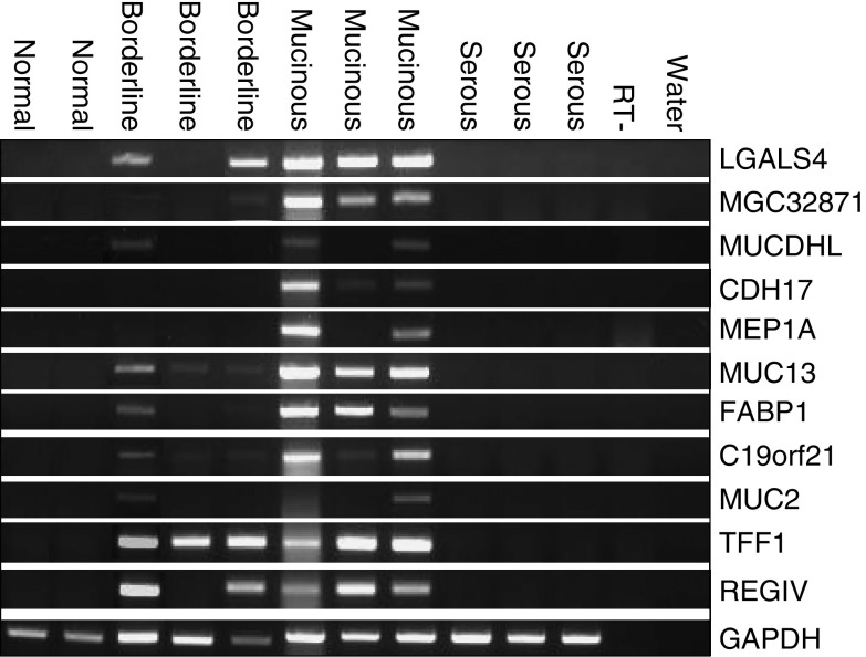
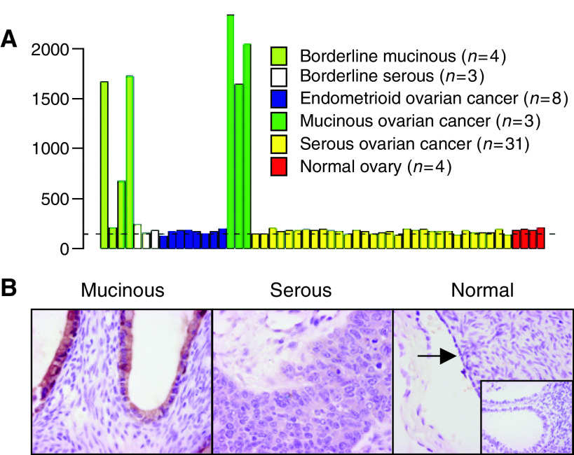
Mucinous epithelial ovarian cancers (MOC) are clinically and morphologically distinct from the other histological subtypes of ovarian cancer. To determine the genetic basis of MOC and to identify potential tumour markers, gene expression profiling of 49 primary ovarian cancers of different histological subtypes was performed using a customised oligonucleotide microarray containing >59 000 probesets. The results show that MOC express a genetic profile that both differs and overlaps with other subtypes of epithelial ovarian cancer. Concordant with its histological phenotype, MOC express genes characteristic of mucinous carcinomas of varying epithelial origin, including intestinal carcinomas. Differences in gene expression between MOC and other histological subtypes of ovarian cancer were confirmed by RT-PCR and/or immunohistochemistry. In particular, galectin 4 (LGALS4) was highly and specifically expressed in MOC, but expressed at lower levels in benign mucinous cysts and borderline (atypical proliferative) tumours, supporting a malignant progression model of MOC. Hence LGALS4 may have application as an early and differential diagnostic marker of MOC.
Figures

References
-
- Balzar M, Winter MJ, de Boer CJ, Litvinov SV (1999) The biology of the 17-1A antigen (Ep-CAM). J Mol Med 77: 699–712 - PubMed
-
- Barker KT, Jackson LE, Crompton MR (1997) BRK tyrosine kinase expression in a high proportion of human breast carcinomas. Oncogene 15: 799–805 - PubMed
-
- Benjamini Y, Yekutieli D (2001) The control of the false discovery rate in multiple hypothesis testing under dependency. Annals Statistics 29: 1165–1188
-
- Byrd JC, Bresalier RS (2004) Mucins and mucin binding proteins in colorectal cancer. Cancer Metastasis Rev 23: 77–99 - PubMed
-
- Casey RC, Oegema Jr TR, Skubitz KM, Pambuccian SE, Grindle SM, Skubitz AP (2003) Cell membrane glycosylation mediates the adhesion, migration, and invasion of ovarian carcinoma cells. Clin Exp Metastasis 20: 143–152 - PubMed
Publication types
MeSH terms
Substances
LinkOut - more resources
Full Text Sources
Other Literature Sources
Medical
