. 2006 Apr;3(4):e100.
doi: 10.1371/journal.pmed.0030100. Epub 2006 Mar 7.

Stage-specific action of matrix metalloproteinases influences progressive hereditary kidney disease

Affiliations

Stage-specific action of matrix metalloproteinases influences progressive hereditary kidney disease

Michael Zeisberg et al. PLoS Med. 2006 Apr.

Abstract

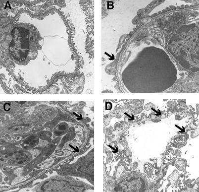
Background: Glomerular basement membrane (GBM), a key component of the blood-filtration apparatus in the in the kidney, is formed through assembly of type IV collagen with laminins, nidogen, and sulfated proteoglycans. Mutations or deletions involving alpha3(IV), alpha4(IV), or alpha5(IV) chains of type IV collagen in the GBM have been identified as the cause for Alport syndrome in humans, a progressive hereditary kidney disease associated with deafness. The pathological mechanisms by which such mutations lead to eventual kidney failure are not completely understood.

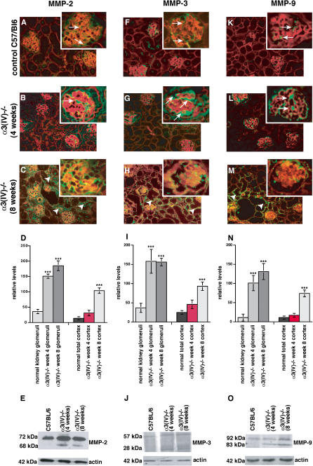
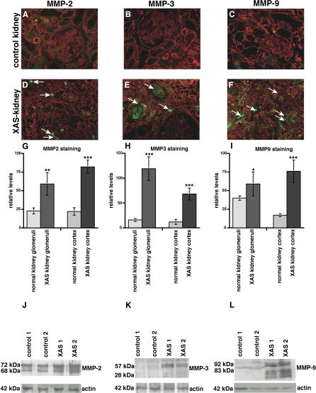
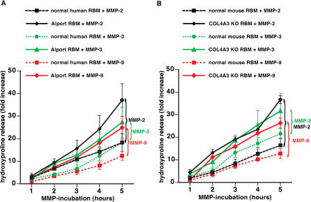
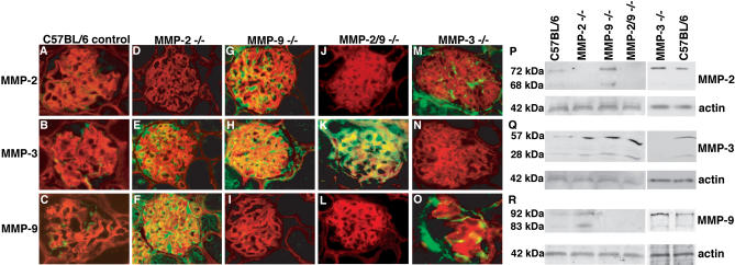
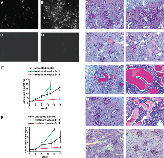
Methods and findings: We showed that increased susceptibility of defective human Alport GBM to proteolytic degradation is mediated by three different matrix metalloproteinases (MMPs)--MMP-2, MMP-3, and MMP-9--which influence the progression of renal dysfunction in alpha3(IV)-/- mice, a model for human Alport syndrome. Genetic ablation of either MMP-2 or MMP-9, or both MMP-2 and MMP-9, led to compensatory up-regulation of other MMPs in the kidney glomerulus. Pharmacological ablation of enzymatic activity associated with multiple GBM-degrading MMPs, before the onset of proteinuria or GBM structural defects in the alpha3(IV)-/- mice, led to significant attenuation in disease progression associated with delayed proteinuria and marked extension in survival. In contrast, inhibition of MMPs after induction of proteinuria led to acceleration of disease associated with extensive interstitial fibrosis and early death of alpha3(IV)-/- mice.

Conclusions: These results suggest that preserving GBM/extracellular matrix integrity before the onset of proteinuria leads to significant disease protection, but if this window of opportunity is lost, MMP-inhibition at the later stages of Alport disease leads to accelerated glomerular and interstitial fibrosis. Our findings identify a crucial dual role for MMPs in the progression of Alport disease in alpha3(IV)-/- mice, with an early pathogenic function and a later protective action. Hence, we propose possible use of MMP-inhibitors as disease-preventive drugs for patients with Alport syndrome with identified genetic defects, before the onset of proteinuria.


Conflict of interest statement

Competing Interests: The authors have declared that no competing interests exist.

Figures

Figure 1
Figure 1. MMP Expression in Kidneys of α3(IV) −/− Mutant and Wild-Type Mice
Frozen sections of control C57/Bl6 mice and α3(IV) −/− mice were stained with primary antibodies for MMP-2, MMP-3, and MMP-9 (FITC-green), and sections were counterstained with Rhodamine-labeled antibodies for entactin (red). (A) In control C57/Bl6 mice, MMP-2 is present sporadically in the glomeruli and in some proximal tubules (arrows). (B) After 4 wk of age, MMP-2 is increased in the glomeruli (arrows) in α3(IV) −/− mice compared to age-matched control kidneys, even before manifestation of proteinuria. (C) After 8 wk of age (after manifestation of GBM-splitting and initiation of interstitial fibrosis), MMP-2 is increased in the interstitium (arrowheads) and in the glomeruli (arrows) in kidneys from α3(IV) −/− mice. (D) MMP-2–staining was quantified using NIH ImageJ software in the glomeruli (left panels) and in total kidney cortex. Glomerular MMP-2–staining was significantly increased at 4 wk in α3(IV) −/− as compared with control. *** p < 0.001 versus control. (E) MMP-2 expression was analyzed by immunoblot using total protein from the kidney cortex of single mice. A representative blot is shown, confirming increased expression of MMP-2 in α3(IV) −/ . The lower blot displays the control blot for actin to confirm equal loading. (F) Immunofluorescence staining revealed that MMP-3–staining is present in the glomeruli (arrows) of control C57/Bl6 mice. (G) MMP-3 is significantly increased in the glomeruli of α3(IV) −/− mice after 4 wk of age. (H) In 8-wk-old α3(IV) −/− mice, MMP-3 is up-regulated in the interstitium (arrowheads) and MMP-3 is also present in the glomeruli (arrows). (I) Quantification of MMP-3–staining revealed a significant increase of MMP-3–staining in the glomeruli and total cortex of α3(IV) −/− mice at 4 wk and 8 wk of age. *** p < 0.001 versus control. (J) Immunoblot analysis revealed increased renal MMP-3 protein. (K) MMP-9 is present in the glomeruli of C57/Bl6 control mice (arrows). (L) The onset of proteinuria is preceded by a significant increase of MMP-9 in the glomeruli of α3(IV) −/− mice (arrows). (M) MMP-9 is significantly up-regulated in the interstitium (arrowheads) and in the glomeruli (arrows) of 8-wk-old α3(IV) −/− mice compared to control. (N) Quantification of immunofluorescence staining using ImageJ software confirmed a significant increase of MMP-9 expression in the glomeruli (left panels) and total cortex region (right panels) of α3(IV) −/−mice as compared with control. *** p < 0.001 versus control. (O) Increased MMP-9 expression was confirmed by immunoblot.
Figure 2
Figure 2. MMP Expression in Kidneys of Patients with XAS and in Normal Human Kidneys
(A–F) Frozen sections of normal human kidneys and kidneys from patients with ESRD due to XAS were stained with antibodies to MMP-2, MMP-3, and MMP-9 (FITC-green), plus laminin (Rhodamine red). Representative stainings of samples obtained from a kidney designated for transplantation or from normal portions of resected kidneys with renal cell carcinoma are displayed. (A–C) Control. (D–F) Kidneys ( n = 2) obtained from male patients with XAS. (G–I) The bar graphs summarize the quantification of MMP-2–staining (G), MMP-3–staining (H), and MMP-9–staining (I). The left bars summarize the evaluation of MMP-staining in the glomeruli; the right bars summarize the MMP-staining in the total kidney cortex in each panel. *** p < 0.001 versus control; ** p < 0.005 versus control; * p < 0.01 versus control. (J–L) Total protein was isolated from normal human kidneys and from two kidneys obtained from two different patients with ESRD due to XAS. Protein was analyzed by SDS-PAGE and immunoblot using specific antibodies to MMP-2 (J), MMP-3 (K), and MMP-9 (L). The lower blots in each panel display the actin control blot to control for equal protein loading.
Figure 3
Figure 3. Increased Susceptibility to Proteolytic Degradation by MMPs of RBM from Patients with XAS and α3(IV) −/− Mice
(A) Degradation of human RBM by MMP-2, MMP-3, and MMP-9. RBM was isolated from human control kidneys and from patients with XAS. RBM was incubated with MMPs, and proteolysis was assessed by estimation of hydroxyproline release. The graph displays the relative increase of hydroxyproline over time. Experiments using normal RBM are represented by dotted lines; studies with RBM from patients with XAS are represented by a full line. Hydroxyproline release after MMP-2 incubation is displayed by black lines, MMP-3 incubation by green lines, and MMP-9 incubation by red lines. Proteolysis of Alport BM was significantly enhanced compared with normal human RBM by each of these MMPS. (B) Degradation of mouse RBM by MMP-2, MMP-3, and MMP-9. RBM was collected from normal C57BL/6 mice ( n = 6) and from α 3(IV) −/− mice ( n = 6) and subjected to active MMPs. Experiments using normal RBM are represented by dotted lines; studies with RBM from α3(IV) −/− mice are represented by a full line. Hydroxyproline release after MMP-2 incubation is displayed by black lines, MMP-3 incubation by green lines, and MMP-9 incubation by red lines. Similarly to human Alport RBM, the RBM from α3(IV) −/− mice displayed an increased susceptibility to proteolysis by MMPs.
Figure 4
Figure 4. Compensation of BM-Degrading MMPs in MMP-2 −/ , MMP-3 −/ , and MMP-9 −/− Mice, and in MMP-2 −/− and MMP-9 −/− Mice
Immunohistochemical staining was performed with MMP-2, MMP-3, or MMP-9–antibodies (green) and with antibodies for entactin (red) on frozen kidney sections from MMP-2 −/− and MMP-9 −/− mice, and from MMP-2 −/− and MMP-9 −/− mice plus C57Bl/6 age-matched control mice. (A–C) Wild-type control kidney. Immunohistochemical staining demonstrated basal expression of MMP-2, MMP-3, or MMP-9 in the glomeruli of C57BL/6 mice. (D–F) MMP-2 −/− kidney. In MMP-2 −/− mice, the absence of MMP-2 was associated with a compensatory increase of MMP-3 and MMP-9. (G–I) MMP-9 −/− kidney. MMP-9–deficiency was associated with increased MMP-2– and MMP-3–staining. (J–L) MMP-2 −/− and MMP-9 −/− kidney. The absence of both MMP-2 and MMP-9 was associated with a substantial increase of MMP-3 in the glomeruli of MMP-2 −/− or MMP-9 −/− double-mutant mice. (M–O) MMP-3 −/− kidney. In MMP-3 −/− mice, the absence of MMP-3 was associated with increased glomerular staining for MMP-2 and MMP-9. (P–R) Immunoblot analysis. Immunoblot analysis of total kidney-cortex protein preparations with antibodies specific for MMP-2 (M), MMP-3 (N), and MMP-9 (O) confirmed compensatory up-regulation of MMPs. The blots were stripped and reprobed with antibodies to actin in order to confirm equal loading (lower panels).
Figure 5
Figure 5. Effect of Combined MMP-Inhibition on the Progression of Renal Disease in α3(IV) −/− Mice
(A–D) In situ zymography. In situ zymography displayed gelatin-degrading activity in normal kidneys (A), which was substantially increased in kidneys of 14-wk-old COL4A3 −/− mice (B). Incubation with the MMP-2, MMP-3, or MMP-9–inhibitor cocktail significantly blocked MMP activities in kidneys from wild-type mice (C) and kidneys from COL4A3 −/− mice (D). (E and F) Renal function in α3(IV) −/− mice. Urinary protein excretion (E) was measured in urine, which was collected over a 24-h period in metabolic cages. Serum creatinine (F) was determined in serum samples that were obtained bi-weekly from mice. The graphs display the progression of urinary protein excretion and serum creatinine levels from 4 to 14 wk of age. In each panel, disease progression is shown in untreated control mice (black), in mice that received MMP-inhibitors starting at 5 wk of age (red), and in mice that received MMP-inhibitors starting at 8 wk of age (green). (G–N) Kidney histology from α3(IV) −/− mice. The images display representative periodic acid-Schiff (PAS)–stained kidney sections from α3(IV) −/− mice, together with images displaying representative kidneys from untreated α 3(IV) −/− mice to illustrate disease progression (G–L). At the age of 3 wk, the kidneys appear completely normal (G). At 4 wk of age, a few glomeruli appear hypercellular, representative of inflammatory cells (H). At the age of 6 wk, a few tubules filled with protein casts appear (I). At 8 wk of age, a few sclerotic glomeruli are present, and atrophic tubules containing protein casts become more abundant (J). At the age of 10 wk, increased numbers of mononuclear cells associated with widening of the interstitial space indicate the onset of interstitial fibrosis (K). At 14 wk of age, the kidneys display severe tubular atrophy, glomerulosclerosis, and interstitial fibrosis, reflecting ESRD (L). A kidney section is displayed of a 14-wk-old α3(IV) −/− mouse that received MMP-inhibitors from 4 wk of age (M). Initiation of treatment after week 4 (compare with H) led to substantially ameliorated disease after 14 wk (M, compare with L). A representative kidney section of a α3(IV) −/− mice at week 11 that received MMP-2–inhibitors, MMP-9–inhibitors, and MMP-3–inhibitors, starting at week 8 and displaying enhanced progression of disease, is displayed (N, compare with K). (Original magnification of PAS-stained histologies: ×200). (O and P) Representative kidney sections from wild-type control mice at 4 wk of age (O) and 14 wk of age (P) are displayed. (Original magnification of PAS-stained histologies: ×200).
Figure 6
Figure 6. Ultra-Structural Analysis (Transmission Electron Microscopy) of GBM from Control and MMP-Inhibitor–Treated Mice
(A) Control C57Bl/6 mice at 14 wk of age. The 5-wk-old normal mice also exhibited normal GBM architecture (unpublished data). (B) Kidneys from 5-wk-old α3(IV) −/− mice before the onset of proteinuria, displaying the beginning of splitting of the GBM and podocyte effacement (arrow). (C). Kidneys from α3(IV) −/− mice at 14 wk of age, which were treated from 5 wk of age with MMP-inhibitors for 9 wk, displayed moderate GBM-splitting and podocyte effacement (arrows). (D) Kidneys from 12-wk-old α3(IV) −/− mice without treatment showed severe lesions of the GBM associated with podocyte effacement (arrows). Original magnification: ×12,250.

Similar articles

Cited by

References

    1. Kalluri R. Basement membranes: Structure, assembly and role in tumour angiogenesis. Nat Rev Cancer. 2003;3:422–433. - PubMed
    1. Timpl R. Structure and biological activity of basement membrane proteins. Eur J Biochem. 1989;180:487–502. - PubMed
    1. Yurchenco PD. Assembly of basement membranes. Ann N Y Acad Sci. 1990;580:195–213. - PubMed
    1. Timpl R, Wiedemann H, van Delden V, Furthmayr H, Kuhn K. A network model for the organization of type IV collagen molecules in basement membranes. Eur J Biochem. 1981;120:203–211. - PubMed
    1. Longo I, Porcedda P, Mari F, Giachino D, Meloni I, et al. COL4A3/COL4A4 mutations: From familial hematuria to autosomal-dominant or recessive Alport syndrome. Kidney Int. 2002;61:1947–1956. - PubMed

Publication types

MeSH terms