. 2006 Jul 1;397(1):179-86.
doi: 10.1042/BJ20051996.

Characterization of different isoforms of the HIF prolyl hydroxylase PHD1 generated by alternative initiation

Affiliations

Characterization of different isoforms of the HIF prolyl hydroxylase PHD1 generated by alternative initiation

Ya-Min Tian et al. Biochem J. .

Abstract

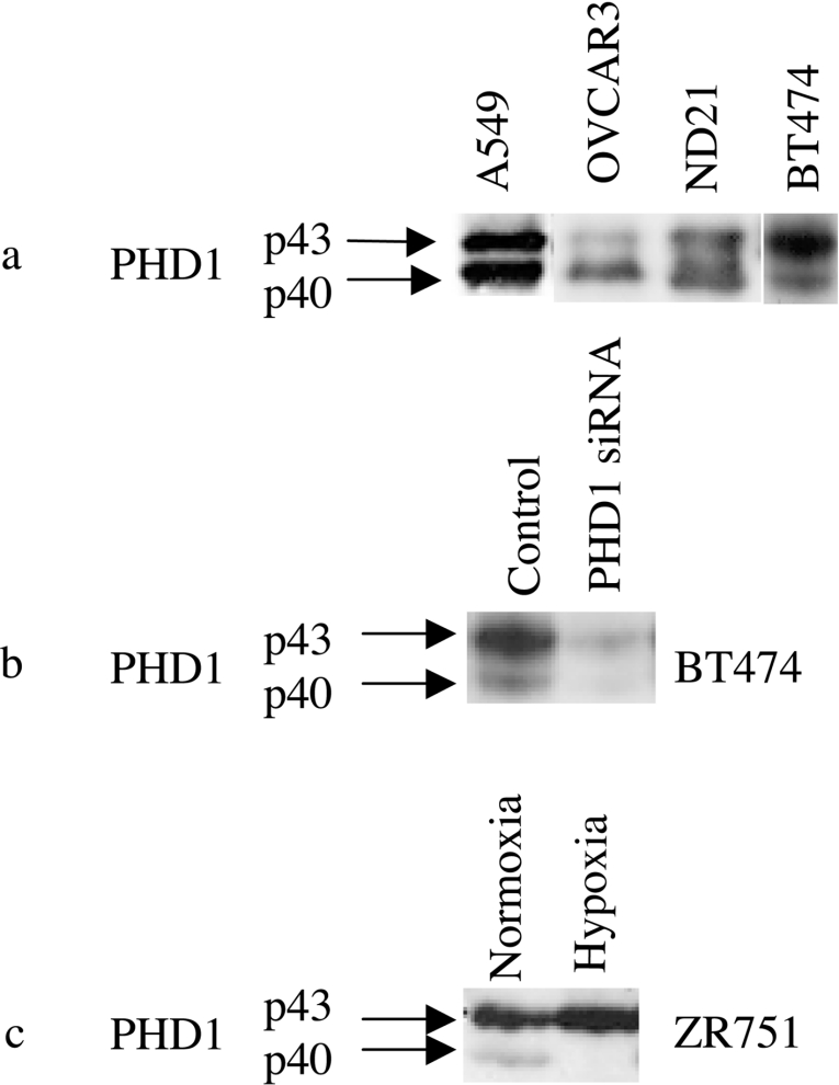
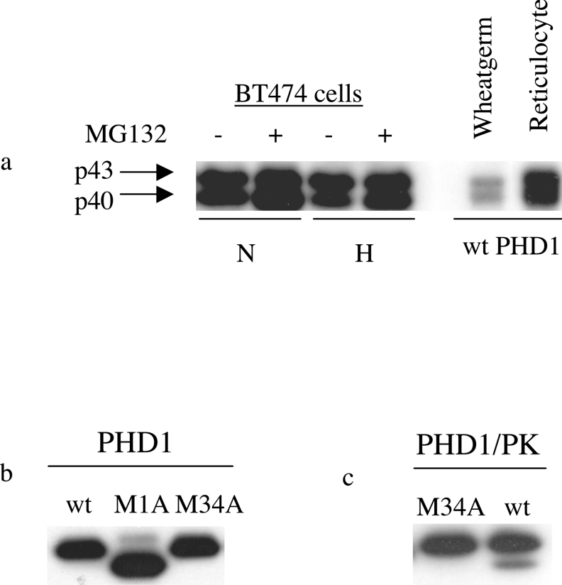
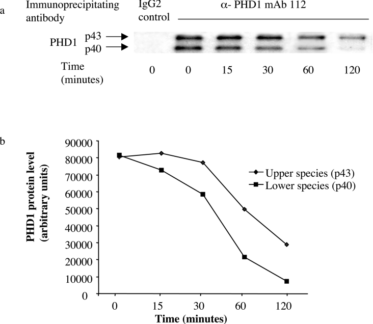
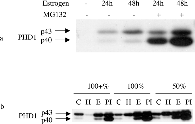
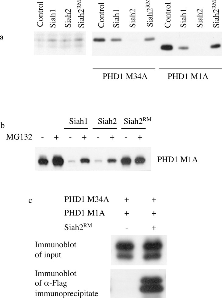
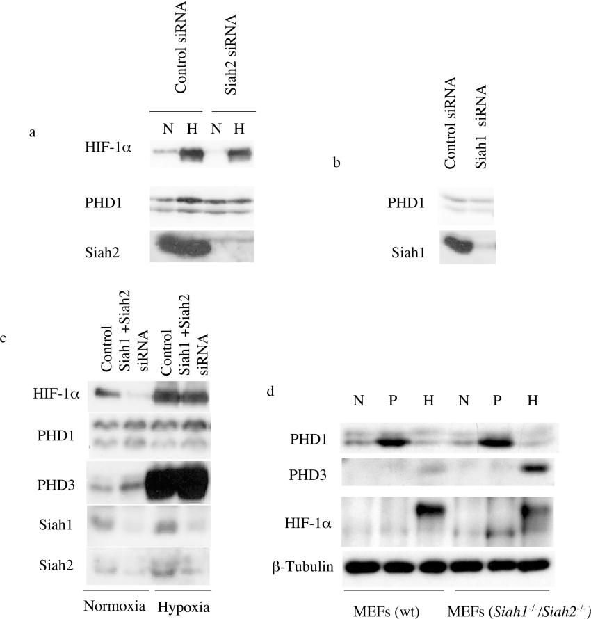
The heterodimeric transcription factor HIF (hypoxia-inducible factor) is central to the regulation of gene expression by oxygen. Three oxygen-dependent prolyl hydroxylase enzymes [PHD1 (prolyl hydroxylase domain 1), PHD2 and PHD3] control the abundance of HIF. In the presence of oxygen, they hydroxylate specific proline residues in HIF-alpha, allowing recognition by pVHL (von Hippel-Lindau protein) and subsequent ubiquitylation and proteasomal destruction. The precise roles and regulation of these enzymes are therefore of particular importance in understanding the physiological and pathological responses to hypoxia. In the present study, we define the existence of two species of PHD1 and provide evidence that they are generated by alternative translational initiation. We demonstrate that these alternative forms are both biologically active with similar HIF prolyl hydroxylase activity but that they differ in their responses to oestrogen, cell confluence and proteasomal inhibition. We show that the two PHD1 species are subject to proteolytic regulation but differ markedly in their protein stability. Though each isoform has the potential to interact with members of the Siah (seven in absentia homologue) ubiquitin ligase family, genetic studies indicated that other proteolytic mechanisms are responsible for control of stability under the conditions examined. The data define the existence of a further level of control in the pathway that regulates cellular responses to hypoxia.


Figures

Figure 1
Figure 1. Expression of two species of PHD1 protein in different cell lines
(a) Immunoblot of cell extracts from A549, OVCAR3, ND21 and BT474 cells detecting PHD1. A doublet of two species labelled p43 and p40 is seen. (b) Immunoblot detecting PHD1 in cell extracts from BT474 cells treated with control and PHD1 siRNA oligonucleotides, showing suppression of both PHD1 species by PHD1 siRNA. (c) Immunoblot detecting PHD1 in cell extracts from ZR751 cells under normoxic conditions and after 16 h of hypoxia (1% O2), showing a reduction in expression of the faster mobility species (p40) under hypoxia.
Figure 2
Figure 2. Endogenous expression of the two forms of PHD1 and comparison with IVTT syntheses of wild-type, PHD1M1A and PHD1M34A proteins
(a) Immunoblot detecting PHD1 in cell extracts from BT474 cells treated with normoxia (21% O2) or hypoxia (1% O2) for 16 h with or without the proteasomal inhibitor MG132 (25 μM) and of IVTT syntheses by rabbit reticulocyte lysate or wheatgerm extract programmed with plasmid encoding wild-type PHD1 (pcDNA3 PHD1). Two protein species are detected with similar mobility in cell extract and IVTT syntheses. The faster mobility species (p40) shows increased levels with proteasomal inhibition but reduced levels under hypoxic conditions. (b) Immunoblot detecting PHD1 in rabbit reticulocyte lysate programmed with plasmids encoding wild-type PHD1 (wt, pcDNA3 PHD1) or mutated PHD1 (M1A, pcDNA3 PHD1M1A or M34A, pcDNA3 PHD1M34A). (c) Immunoblot detecting PK in rabbit reticulocyte lysate programmed with plasmids encoding wild-type PHD1 with a C-terminal PK epitope tag (pcDNA3 PHD1PK) and a mutated version (pcDNA3 PHD1M34APK).
Figure 3
Figure 3. Pulse–chase study examining for a precursor–product relationship between the two species of PHD1
(a) Pulse–chase radiolabelling studies of PHD1. ZR751 cells were treated with [35S]methionine/cysteine for 30 min followed by incubation for 0, 15, 30, 60 or 120 min and immunoprecipitation of cell extract was then performed with the anti-PHD1 antibody mAb 112 or a control antibody. The immunoprecipitates were separated by SDS/PAGE, and 35S signals of the upper (p43) and lower (p40) species were quantified by a phosphoimager screen and are displayed graphically in (b).
Figure 4
Figure 4. Expression of the two species of PHD1 in response to oestrogen, proteasomal inhibition, hypoxia and cellular confluence
(a) Immunoblot detecting the two species of PHD1 (p43 and p40) in cell extracts from ZR751 cells treated with oestrogen (10 ng/ml oestradiol-17β) for 24 or 48 h as indicated and/or the proteasomal inhibitor MG132 (25 μM) for the last 4 h. (b) Immunoblot detecting the two species of PHD1 (p43 and p40) in cell extracts from ZR751 cells cultured at varying levels of confluence (50%, 100% and 100%+) and treated with hypoxia (‘H’; 1% O2) for 16 h, oestrogen (‘E’; 10 ng/ml oestradiol-17β) for 48 h, or oestrogen for 48 h together with proteasomal inhibition for the last 4 h of incubation (‘PI’; 25 μM MG132). ‘C’, control.
Figure 5
Figure 5. Cycloheximide chase examining the protein stability of the two species of PHD1 (p43 and p40) under hypoxic and normoxic conditions
U2OS cells were transfected with plasmids encoding wild-type or mutant versions of PHD1 (Wt, pcDNA3 PHD1; M1A, pcDNA3 PHD1M1A; or M34A, pcDNA3 PHD1M34A) or no plasmid and treated with normoxia (21% O2) or hypoxia (1% O2) for 16 h prior to treatment with 0.1 mM cycloheximide for 0, 30, 60 or 90 min. Endogenous and transfected protein levels of PHD1 and PHD3 were detected by immunoblotting.
Figure 6
Figure 6. Prolyl hydroxylase activity of wild-type, PHD1M1A and PHD1M34A enzymes
Proteins synthesized in rabbit reticulocyte lysate were assayed for their ability to convert a HIF-1α peptide (HIF-1α residues 556–574) into a VHL-binding form. PHD1 enzymes were produced in a rabbit reticulocyte lysate IVTT and relative abundance of each protein was quantified by SDS/PAGE and autoradiography using a phosphoimager and equimolar amounts of the enzymes were then used in the assay. The N-terminal biotinylated peptide corresponding to human HIF-1α residues 556–574 was treated with the enzymes and subsequent binding to 35S-radiolabelled VHL protein was quantified by SDS/PAGE and autoradiography. Reactions were performed in triplicate and results are given as means±S.D. No statistically significant differences were seen between the enzymatic activities of the different forms of PHD1.
Figure 7
Figure 7. Effect of PHD1 expression on the activity of Gal4 fusion proteins bearing isolated HIF-α degradation domains
MCF7 cells were co-transfected with plasmids expressing (i) a Gal4 reporter gene, (ii) β-galactosidase, (iii) the indicated HIF-α ODD (oxygen-dependent degradation domain) sequence as a Gal4–HIF–VP16 fusion protein [HIF-1α/NODD (amino acids 344–553), HIF-1α/CODD-(554–698), HIF-2α/NODD-(345–517) or HIF-2α/CODD-(517–682)] and (iv) wild-type PHD1 (wt, pcDNA3 PHD1) or a mutated version of PHD1 (M1A, pcDNA3 PHD1M1A) as indicated. Luciferase activities were analysed and corrected for transfection efficiency by β-galactosidase activity. Immunoblot analysis of expressed PHD1 protein levels are shown. Experiments were performed in triplicate and results are given as means±S.D. No statistically significant differences were seen between the actions of the different forms of PHD1.
Figure 8
Figure 8. Effect of the Siah E3 ligase overexpression on PHD1 abundance
(a) Plasmids encoding PHD1M1A (pcDNA3 PHD1M1A; 1600 ng) or PHD1M34A (pcDNA3 PHD1M34A; 100 ng) were transfected into MCF7 cells with FLAG-tagged, Siah-encoding expression plasmids (pcDNA3-FLAG-Siah1, pcDNA3-FLAG-Siah2 or pcDNA3-FLAG-Siah2Rm; 100 ng). The abundance of the different forms of endogenous and transfected PHD1 was detected by immunoblotting. Siah2 (and to a lesser extent Siah1) decreased the protein levels of both forms of transfected PHD1. (b) Similar experiments were undertaken to examine effects of Siah expression on PHD1M1A in the absence and presence of the proteasomal inhibitor, MG132, in U2OS cells. (c) HEK-293T cells were co-transfected with plasmids encoding PHD1M1A (pcDNA3 PHD1M1A) or PHD1M34A (pcDNA3 PHD1M34A) and with or without plasmid encoding FLAG–Siah2Rm (pcDNA3-FLAG-Siah2Rm). A specific association was seen between Siah2Rm and PHD1. However, the slower mobility species of PHD1 was predominant in the input, while the faster mobility form dominated in the immunoprecipitate.
Figure 9
Figure 9. Effect of Siah suppression or deficiency on PHD1 expression
MCF7 cells were treated with Siah-targeted siRNA oligonucleotides to suppress the level of Siah1 (a), Siah2 (b) or both Siah1 and Siah2 (c) and extracts were examined for PHD1, HIF-1α, PHD3 and Siah protein expression as indicated. (d) MEFs derived from Siah1a−/−/Siah2−/− or wild-type (wt) mice were treated with normoxia (‘N’; 21% O2), proteasomal inhibition (‘P’; 25 μM MG132) or hypoxia (‘H’; 5% O2) for 5 h. Cell extracts were examined for PHD1, HIF-1α, PHD3 and β-tubulin expression by immunoblotting.

References

    1. Kaelin W. G. J. How oxygen makes its presence felt. Genes Dev. 2002;16:1441–1445. - PubMed
    1. Wang G. L., Semenza G. L. Purification and characterisation of hypoxiainducible factor 1. J. Biol. Chem. 1995;270:1230–1237. - PubMed
    1. Maxwell P. H., Wiesener M. S., Chang G.-W., Clifford S. C., Vaux E. C., Cockman M. E., Wykoff C. C., Pugh C. W., Maher E. R., Ratcliffe P. J. The tumour suppressor protein VHL targets hypoxia-inducible factors for oxygen-dependent proteolysis. Nature (London) 1999;399:271–275. - PubMed
    1. Ivan M., Kondo K., Yang H., Kim W., Valiando J., Ohh M., Salic A., Asara J. M., Lane W. S., Kaelin W. G. J. HIFα targeted for VHL-mediated destruction by proline hydroxylation: implications for O2 sensing. Science. 2001;292:464–468. - PubMed
    1. Jaakkola P., Mole D. R., Tian Y.-M., Wilson M. I., Gielbert J., Gaskell S. J., Kriegsheim A. V., Hebestreit H. F., Mukherji M., Schofield C. J., et al. Targeting of HIF-α to the von Hippel-Lindau ubiquitylation complex by O2-regulated prolyl hydroxylation. Science. 2001;292:468–472. - PubMed

Publication types