. 2006 May;17(5):2287-302.
doi: 10.1091/mbc.e05-11-1089. Epub 2006 Mar 1.

Coordinated requirements of human topo II and cohesin for metaphase centromere alignment under Mad2-dependent spindle checkpoint surveillance

Affiliations

Coordinated requirements of human topo II and cohesin for metaphase centromere alignment under Mad2-dependent spindle checkpoint surveillance

Yusuke Toyoda et al. Mol Biol Cell. 2006 May.

Abstract

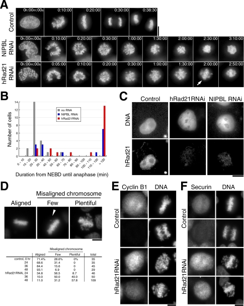
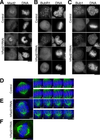
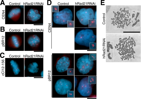
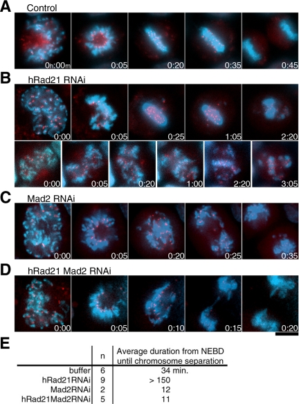
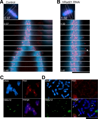
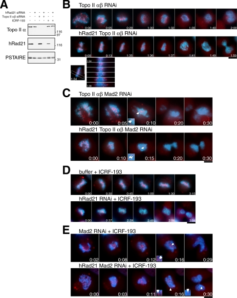
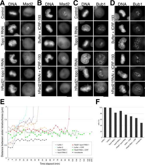
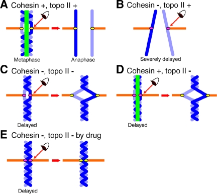
Cohesin maintains sister chromatid cohesion until its Rad21/Scc1/Mcd1 is cleaved by separase during anaphase. DNA topoisomerase II (topo II) maintains the proper topology of chromatid DNAs and is essential for chromosome segregation. Here we report direct observations of mitotic progression in individual HeLa cells after functional disruptions of hRad21, NIPBL, a loading factor for hRad21, and topo II alpha,beta by RNAi and a topo II inhibitor, ICRF-193. Mitosis is delayed in a Mad2-dependent manner after disruption of either or both cohesin and topo II. In hRad21 depletion, interphase pericentric architecture becomes aberrant, and anaphase is virtually permanently delayed as preseparated chromosomes are misaligned on the metaphase spindle. Topo II disruption perturbs centromere organization leading to intense Bub1, but no Mad2, on kinetochores and sustains a Mad2-dependent delay in anaphase onset with persisting securin. Thus topo II impinges upon centromere/kinetochore function. Disruption of topo II by RNAi or ICRF-193 overrides the mitotic delay induced by cohesin depletion: sister centromeres are aligned and anaphase spindle movements occur. The ensuing accumulation of catenations in preseparated sister chromatids may overcome the reduced tension arising from cohesin depletion, causing the override. Cohesin and topo II have distinct, yet coordinated functions in metaphase alignment.


Figures

Figure 1.
Figure 1.
hRad21 and NIPBL knockdown cells are mitotically delayed without securin destruction. (A) Time-lapse images taken from movies (Supplement 2) are shown. Top, control no RNAi; middle, NIPBL RNAi (120 h after RNAi); bottom, hRad21 RNAi (104 h after RNAi). Arrow, the partially formed plate. (B) The duration from NEBD until the onset of anaphase was measured in the movies of control non-RNAi, NIPBL, and hRad21 RNAi cells. (C) hRad21 retention in nuclei. Transfected cells were extracted in CSK buffer supplemented with Triton X-100 before fixation. Cells shown are 96 h after transfection. Top, DNA stained with Hoechst 33342; bottom, immunostaining of hRad21. (D) Top, left, normal metaphase; middle, cell showing a few misaligned chromosomes (arrowhead); right, cell showing abundant misaligned chromosomes. Bottom, the frequencies of each appearance measured in control non-RNAi and hRad21 RNAi cells. (E and F) Securin and cyclin B1 were immunostained in control non-RNAi and hRad21 RNAi cells fixed by paraformaldehyde. In hRad21 RNAi cells, mitotic cells containing abundant EMCs are shown. Bars, 10 μm.
Figure 2.
Figure 2.
Bub1, BubR1-positive, and Mad2-negative kinetochores in hRad21 knockdown cells are end-on associated with microtubules. (A–C) Mitotic cells of control non-RNAi and hRad21 RNAi (48 h) cells were stained for DNA with Hoechst 33342, Mad2 (A), BubR1 (B), and Bub1 (C) with antibodies. (D and E) Eight serial sections (0.4-μm intervals) along the z-axis were taken for metaphase in control non-RNAi cells (D) and for hRad21 RNAi cell (E). Red, anti-CENP-C; green, tubulin; blue, DNA. (F) A hRad21 RNAi cell that retained a plate-like structure. Bars, 10 μm.
Figure 3.
Figure 3.
Mitotic and interphase DNAs show aberrant behavior and organization. (A–C) FISH was used for mitotic cells of control non-RNAi and after hRad21 RNAi, using chromosome specific peri-centromeric probes (A and B for chromosomes 4 and 12, respectively) and peri-telomeric probe (C). (D) FISH was applied for interphase cells. (E) Chromosomes were spread and Giemsa stained. Top, control no RNAi; bottom, hRad21 RNAi cells (48 h). Bars, 10 μm.
Figure 4.
Figure 4.
Sister kinetochore movements are under the Mad2 surveillance. (A–D) A number of movies were taken, and example micrographs are shown. Red, hMis12-GFP; blue, DNA. Elapsed time was shown as h:min. Bar, 10 μm. (A) Control cell. (B) hRad21 RNAi cells. Top, cell displaying a few EMCs. Bottom, cell showing abundant EMCs. No anaphase-like centromere segregation was observed. (C) Mad2 RNAi cell. (D) hRad21 Mad2 double RNAi cell. (E) The average duration (min) from NEBD to the onset of anaphase.
Figure 5.
Figure 5.
Preseparated centromere signals are under the tension but do not contain detectable hRad21. (A and B) Movie images taken for control non-RNAi (A) and hRad21 RNAi (B) cells are selected for the same chromosome area (indicated with the frame in the small top micrographs) and arranged in a time-lapse order (1-min intervals). GFP-hMis12 signals (red) and chromosome DNA (blue). Elapsed time was shown as h:min. The arrowhead indicates a sister centromere signal. (C and D) Chromosome spread of control non-RNAi (C) and hRad21 RNAi (D). Blue, DNA; red, Myc-tagged hRad21; green, hMis12. Arrows, three remotely paired chromatids. Spread and staining conditions were aimed to maintain the intact structure. Bars, 10 μm.
Figure 6.
Figure 6.
Defect in topo II induces the Mad2-dependent delay, abolishes EMCs, and causes the tension. (A) Immunoblot of HeLa cell extracts with or without RNAi for topo II α,β and/or hRad21 using antibodies against topo IIα and hRad21. PSTAIRE antibody against Cdc2 was used as a loading control. (B) Time-lapse images of cells (red, hMis12; blue, DNA) after the topo IIα,β double RNAi (top) and the topo IIα,β hRad21 triple RNAi (middle). Time-lapse images for the concerted centromere segregation in the triple RNAi (bottom). (C) Time-lapse images of cells after the topo IIα,β Mad2 triple RNAi (top) and the topo IIα,β hRad21 Mad2 quadruple RNAi (bottom). Enlarged insets were shown to indicate apparent sister separation in comparison with persistent pairing shown in insets in E. Arrowheads, the GFP-hMis12 signals that were considered as sister kinetochores. (D) Movie images of cells treated with the ICRF-193 without or with knockdown of hRad21 (top and bottom, respectively). (E) Time-lapse images of Mad2 single (top) or hRad21 Mad2 double (bottom) RNAi cells treated with ICRF-193. Note that in both cases the pairing of sister kinetochores persisted when the GFP-hMis12 signals started to diminish as shown in insets. Bars, 10 μm.
Figure 7.
Figure 7.
The mode of microtubule–kinetochore association in topo II–defective cells. (A–D) Kinetochore localization of Mad2 and Bub1 in topo II–deficient cells. Cells treated as indicated were fixed with paraformaldehyde and immunostained by anti-Mad2 (A and B) or anti-Bub1 (C and D) antibodies. Transfected cells were harvested after 48 h with or without an additional incubation for 2 h in the presence of ICRF-193. In each treatment, Mad2 and Bub1 were confirmed to localize in prometaphase or prophase chromosomes, respectively. Bar, 10 μm. (E) Based on movie data, the distances between discernible sister kinetochores were measured and plotted on the graph. Data obtained from nocodazole-treated cells served as the control where no spindle and tension existed. (F) The average distances between discernible sister kinetochores during the metaphase-like stage are shown. The value of buffer-transfected control (buffer 1; 1.45 μm) was set to 100%.
Figure 8.
Figure 8.
A model: how cohesin and topo II are implicated in metaphase chromosome alignment. A model illustrating how cohesin and topo II contribute to chromosome alignment in metaphase. (A) Metaphase and anaphase chromosomes of non-RNAi HeLa cells. (B) Preseparated chromosomes in hRad21RNAi. (C) hRad21 Topo II RNAi cells. (D) Topo II RNAi cells. (E) hRad21 RNAi cells treated with ICRF-193. Chromosome, blue; the link regulated by cohesin and topo II, green; spindle, orange. The eye represents surveillance by Mad2-dependent spindle checkpoint control. Kinetochores colored in pink indicate activation of spindle checkpoint, whereas the inactivated checkpoint is indicated by yellow kinetochores. Spindle checkpoint is activated in all the cases except for control non-RNAi cells.

References

    1. Atienza, J. M., Roth, R. B., Rosette, C., Smylie, K. J., Kammerer, S., Rehbock, J., Ekblom, J., and Denissenko, M. F. (2005). Suppression of RAD21 gene expression decreases cell growth and enhances cytotoxicity of etoposide and bleomycin in human breast cancer cells. Mol. Cancer Ther. 4, 361–368. - PubMed
    1. Baldini, A., Rocchi, M., Archidiacono, N., Miller, O. J., and Miller, D. A. (1990). A human alpha satellite DNA subset specific for chromosome 12. Am. J. Hum. Genet. 46, 784–788. - PMC - PubMed
    1. Bhalla, N., Biggins, S., and Murray, A. W. (2002). Mutation of YCS4, a budding yeast condensin subunit, affects mitotic and nonmitotic chromosome behavior. Mol. Biol. Cell 13, 632–645. - PMC - PubMed
    1. Biggins, S., and Murray, A. W. (2001). The budding yeast protein kinase Ipl1/Aurora allows the absence of tension to activate the spindle checkpoint. Genes Dev. 15, 3118–3129. - PMC - PubMed
    1. Carpenter, A. J., and Porter, A. C. (2004). Construction, characterization, and complementation of a conditional-lethal DNA topoisomerase IIalpha mutant human cell line. Mol. Biol. Cell 15, 5700–5711. - PMC - PubMed

Publication types

MeSH terms

Substances