. 2006 Mar 1;34(4):1293-303.
doi: 10.1093/nar/gkl019. Print 2006.

The hepatitis C virus 3'-untranslated region or a poly(A) tract promote efficient translation subsequent to the initiation phase

Affiliations

The hepatitis C virus 3'-untranslated region or a poly(A) tract promote efficient translation subsequent to the initiation phase

Shelton S Bradrick et al. Nucleic Acids Res. .

Abstract

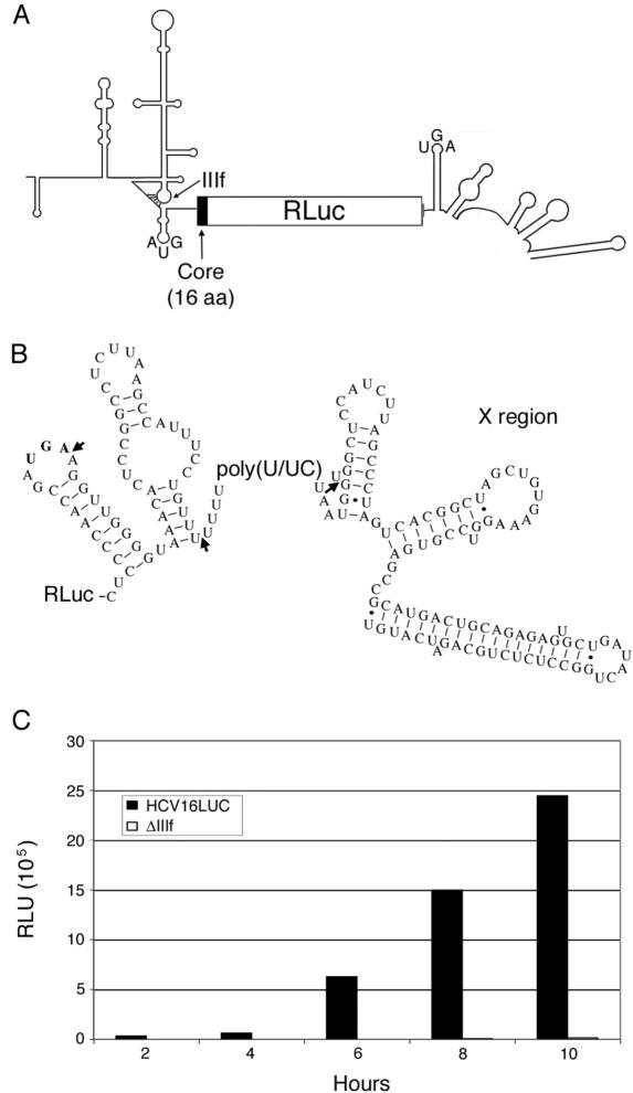
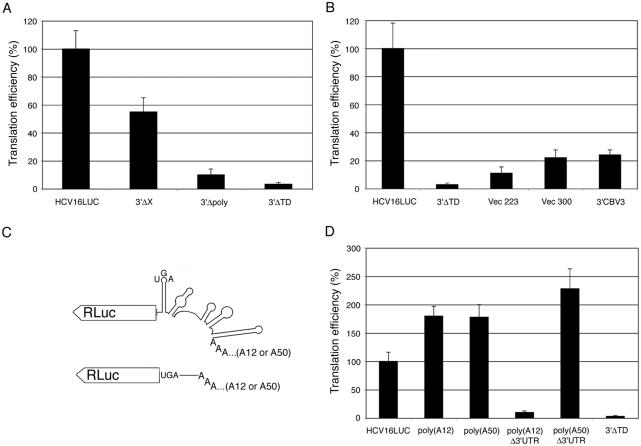
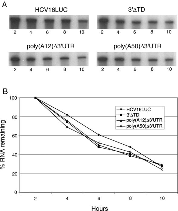
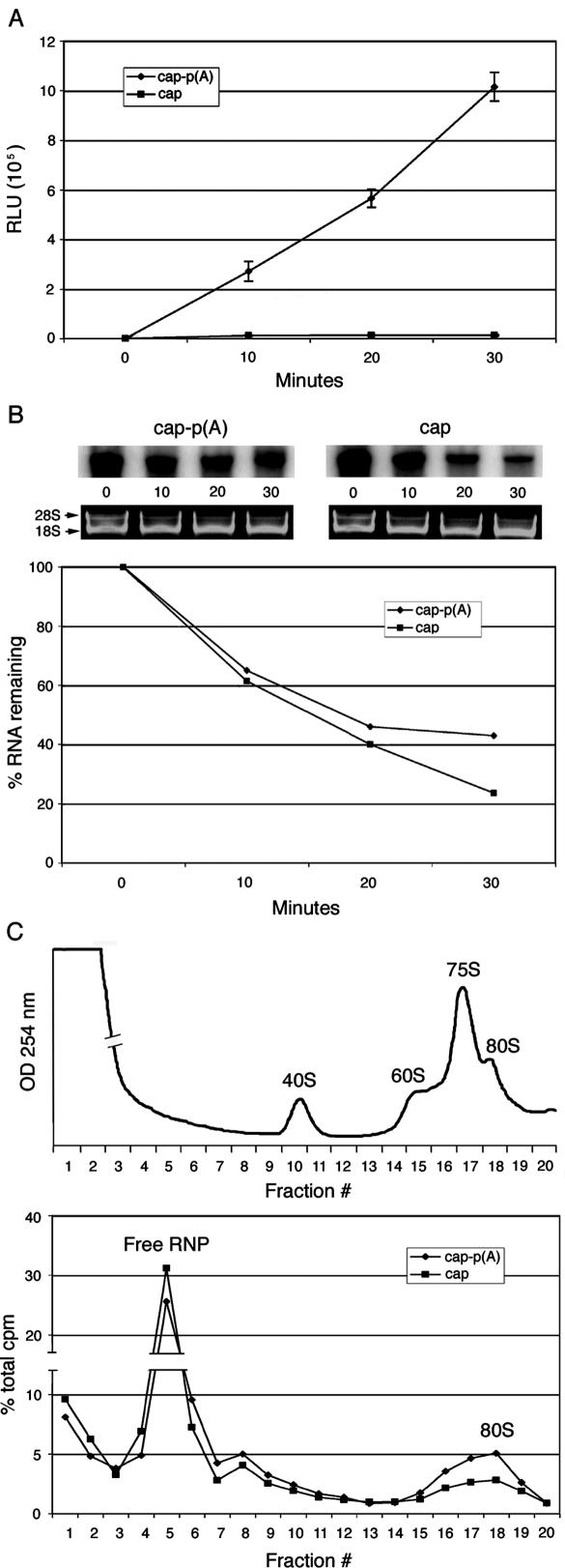
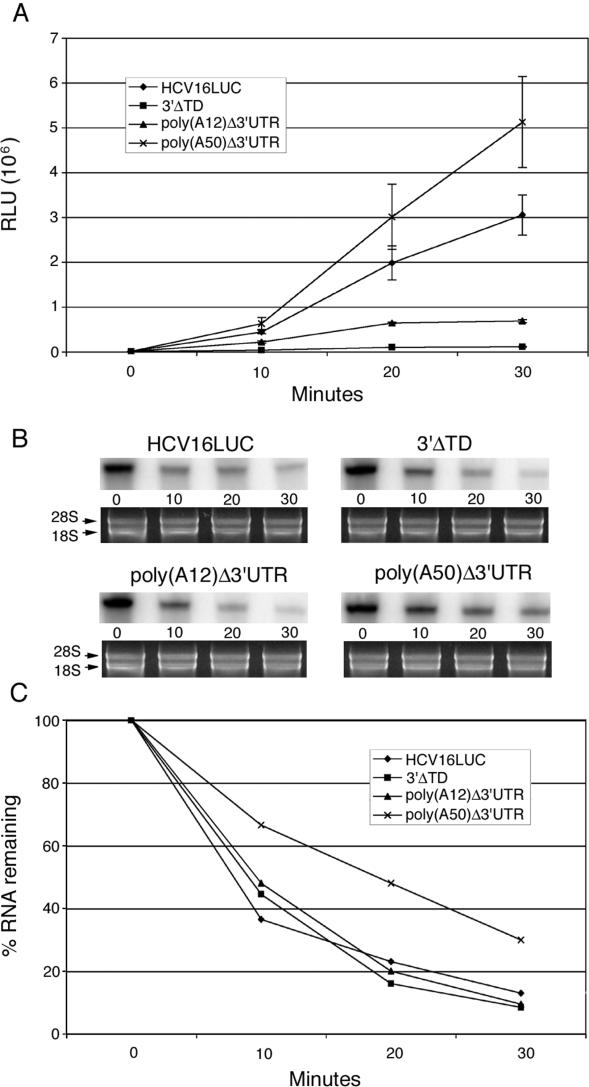
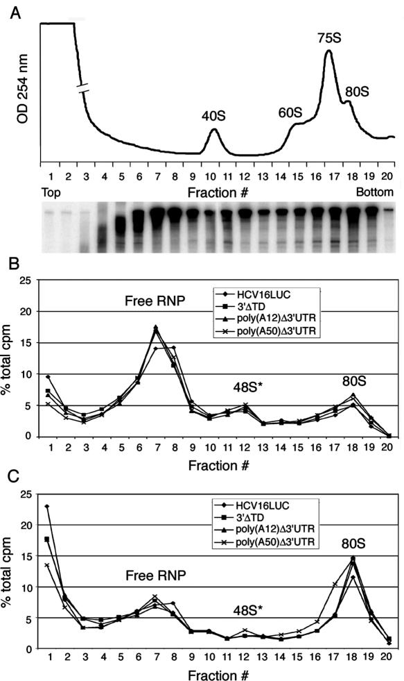
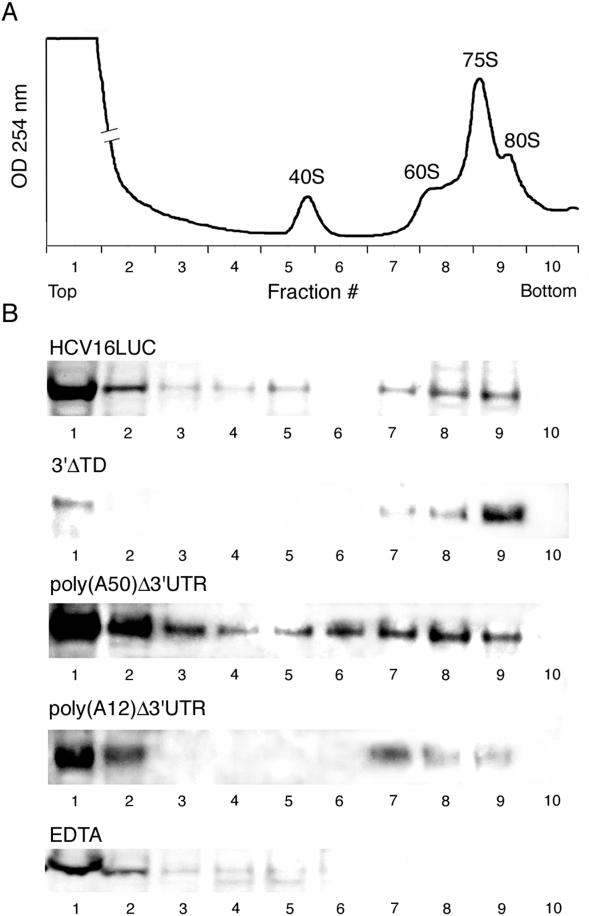
Enhancement of eukaryotic messenger RNA (mRNA) translation initiation by the 3' poly(A) tail is mediated through interaction of poly(A)-binding protein with eukaryotic initiation factor (eIF) 4G, bridging the 5' terminal cap structure. In contrast to cellular mRNA, translation of the uncapped, non-polyadenylated hepatitis C virus (HCV) genome occurs independently of eIF4G and a role for 3'-untranslated sequences in modifying HCV gene expression is controversial. Utilizing cell-based and in vitro translation assays, we show that the HCV 3'-untranslated region (UTR) or a 3' poly(A) tract of sufficient length interchangeably stimulate translation dependent upon the HCV internal ribosomal entry site (IRES). However, in contrast to cap-dependent translation, the rate of initiation at the HCV IRES was unaffected by 3'-untranslated sequences. Analysis of post-initiation events revealed that the 3' poly(A) tract and HCV 3'-UTR improve translation efficiency by enabling termination and possibly ribosome recycling for successive rounds of translation.


Figures

Figure 1
Figure 1
Efficient translation of subgenomic HCV reporter RNA depends upon higher order RNA structure. (A) Schematic representation of HCV16LUC reporter RNA. The positions of IRES domain IIIf and conserved pseudoknot are indicated. (B) Sequence and secondary structure of the HCV 3′-UTR. Arrows indicate locations of 3′ terminal deletions assayed for translation efficiency in Huh7 cells (Figure 2). The authentic stop codon is indicated in bold letters. (C) Huh7 cells were transfected with 0.2 µg of indicated reporter RNAs and RLuc was measured by enzymatic assay over the course of 10 h after the start of transfection.
Figure 2
Figure 2
HCV IRES-dependent translation is stimulated by the cognate HCV 3′-UTR and artificial polyadenylation. Huh7 cells were co-transfected with the indicated reporter RNA and a second mRNA encoding firefly luciferase to control for variability of transfection efficiency. Six hours post-transfection, cell lysates were harvested for determination of luciferase activities. Translation efficiency of intact HCV16LUC was arbitrarily set at 100%. Error bars indicate standard deviation of three independently performed experiments. Effects of (A) progressive HCV 3′-UTR deletion, (B) substitution of the HCV 3′-UTR with non-specific sequences and (D) 3′ polyadenylation on RLuc expression are shown. A representation of the 3′ region of polyadenylated RNA constructs is illustrated in (C).
Figure 3
Figure 3
Stability of HCV reporter RNAs is unaffected by 3′-UTR modification in vivo. (A) Radioactively labeled reporter RNAs were transfected into Huh7 cells. At the indicated time points (hours) after transfection, cells were lysed for isolation of soluble cytoplasmic RNA (see Materials and Methods). Extracted RNAs were separated by denaturing PAGE and visualized by autoradiography. (B) Intensities of radiolabeled RNA bands were quantitated using a phophorimager. RNA levels at 2 h after the start of transfection were set to 100%.
Figure 4
Figure 4
Polyadenyation stimulates cap-dependent translation at the level of initiation in a HeLa cell extract. (A) Extracts were programmed with 150 ng of either cap- or cap-poly(A) mRNA containing the β-globin leader sequence. Aliquots were removed at periodic intervals over the course of a 30 min incubation for measurements of luciferase activity. Error bars indicate standard deviation of reactions performed in triplicate. (B) Internally 32P-labeled capped mRNAs were used to program in vitro translation reactions and decay was monitored over 30 min. Phophorimager analysis of band intensities is displayed. As a control for extraction and precipitation procedures, ribosomal RNAs (28S and 18S) from corresponding samples were visualized by native agarose gel electrophoresis. (C) Radiolabeled capped transcripts were assessed for their ability to incorporate into active 80S ribosomal complexes. Reactions were performed in the presence of 0.5 mM cycloheximide to prevent elongation. After 15 min of incubation at 37oC, reactions were diluted in chilled gradient buffer and loaded onto linear 5–20% sucrose gradients. Twenty fractions were collected from each gradient and analyzed by liquid scintillation counting. An absorbance profile indicating positions of ribosomal subunits and intact ribosomes is shown in the top panel.
Figure 5
Figure 5
HCV IRES-driven translation is stimulated in vitro by poly(A) and the HCV 3′-UTR. In vitro translation reactions were programmed with the indicated uncapped HCV reporter RNAs for analysis of translation efficiency (A) and RNA stability (B and C) over the course of 30 min. Error bars indicate standard deviation. Ribosomal RNAs from corresponding samples were visualized by native agarose gel electrophoresis.
Figure 6
Figure 6
Ability of the HCV IRES to mediate the initiation phase of translation is unaffected by 3′-untranslated sequences. (A) The absorbance profile of a fractionated in vitro translation reaction is shown with positions of free ribosomal subunits and intact ribosomes indicated. The 75S peak represents mRNA-independent association of small and large ribosomal subunits (13). The lower panel shows the distribution of intact poly(A50)Δ3′-UTR reporter RNA at 2 min after start of the reaction by denaturing PAGE. (B) In vitro translation reactions were assembled in the presence of 0.5 mM cycloheximide and allowed to proceed for 2 min before addition of ice-cold gradient buffer to stop the reaction. Reactions were then subjected to sucrose density gradient centrifugation followed by fractionation. Individual fractions were analyzed for radioactivity levels by scintillation counting. (C) Same analysis as in (B) after 5 min of incubation at 37oC.
Figure 7
Figure 7
Analysis of translation termination in vitro by density gradient fractionation and immunoblot. (A) The absorbance profile of a fractionated in vitro translation reaction is shown with positions of free ribosomal subunits and intact ribosomes indicated. (B) In vitro translation reactions programmed with the indicated RNAs and incubated 30 min were fractionated by density gradient centrifugation. Individual fractions were TCA precipitated and separated by SDS–PAGE for western blot using α-RLuc antibody. As a control, an in vitro translation reaction programmed with HCV16LUC RNA was fractionated in the presence of 10 mM EDTA to disrupt association of ribosomal subunits, releasing RLuc to upper fractions of the gradient.

References

    1. Hershey J.W.B., Merrick W.C. The pathway and mechanism of initiation of protein synthesis. In: Sonenberg N., Hershey J.W.B., Mathews M.B., editors. Translational Control of Gene Expression. Cold Spring Harbor, NY: Cold Spring Harbor Laboratory Press; 2000. pp. 33–88.
    1. Mangus D.A., Evans M.C., Jacobson A. Poly(A)-binding proteins: multifunctional scaffolds for the post-transcriptional control of gene expression. Genome Biol. 2003;4:223. - PMC - PubMed
    1. Tarun S.Z., Sachs A.B. Association of the yeast poly(A) tail binding protein with translation initiation factor eIF-4G. EMBO J. 1996;15:7168–7177. - PMC - PubMed
    1. Kahvejian A., Svitkin Y.V., Sukarieh R., M'Boutchou M.N., Sonenberg N. Mammalian poly(A)-binding protein is a eukaryotic translation initiation factor, which acts via multiple mechanisms. Genes Dev. 2005;19:104–113. - PMC - PubMed
    1. Searfoss A., Dever T.E., Wickner R. Linking the 3′ poly(A) tail to the subunit joining step of translation initiation: relations of Pab1p, eukaryotic translation initiation factor 5B (Fun12p), and Ski2p-Slh1p. Mol. Cell Biol. 2001;21:4900–4908. - PMC - PubMed

Publication types