Comparative Study
. 2006 Mar 1;20(5):624-36.
doi: 10.1101/gad.1397906.

Specific functional interactions of nucleotides at key -3 and +4 positions flanking the initiation codon with components of the mammalian 48S translation initiation complex

Affiliations
Comparative Study

Specific functional interactions of nucleotides at key -3 and +4 positions flanking the initiation codon with components of the mammalian 48S translation initiation complex

Andrey V Pisarev et al. Genes Dev. .

Abstract

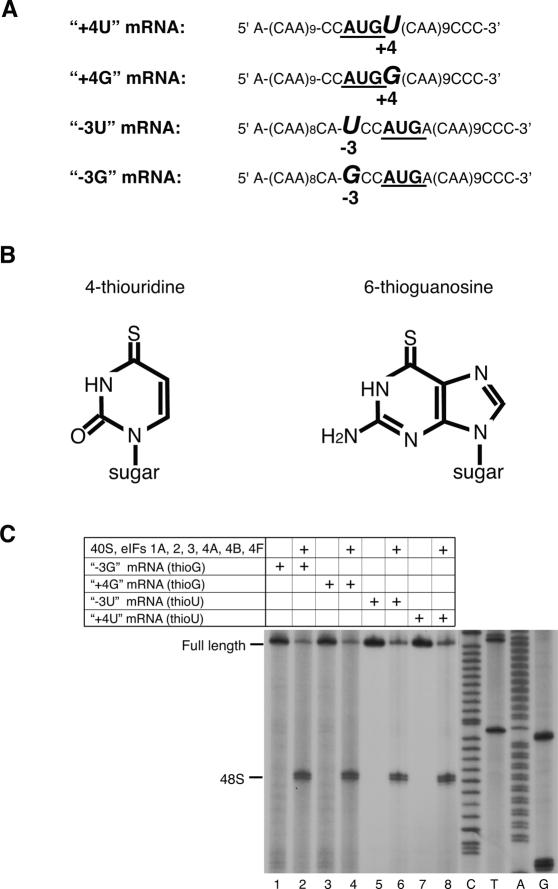
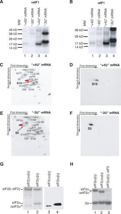
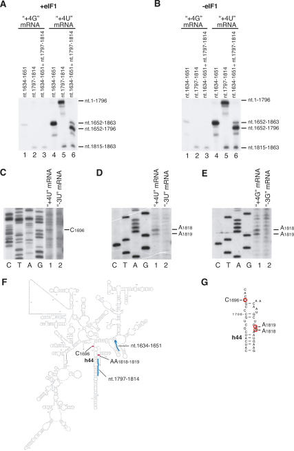
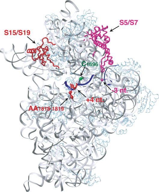
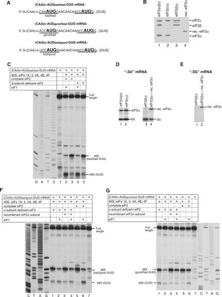
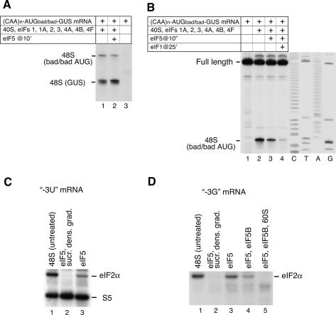
Eukaryotic initiation factor (eIF) 1 maintains the fidelity of initiation codon selection and enables mammalian 43S preinitiation complexes to discriminate against AUG codons with a context that deviates from the optimum sequence GCC(A/G)CCAUGG, in which the purines at (-)3 and (+)4 positions are most important. We hypothesize that eIF1 acts by antagonizing conformational changes that occur in ribosomal complexes upon codon-anticodon base-pairing during 48S initiation complex formation, and that the role of (-)3 and (+)4 context nucleotides is to stabilize these changes by interacting with components of this complex. Here we report that U and G at (+)4 both UV-cross-linked to ribosomal protein (rp) S15 in 48S complexes. However, whereas U cross-linked strongly to C(1696) and less well to AA(1818-1819) in helix 44 of 18S rRNA, G cross-linked exclusively to AA(1818-1819). U at (-)3 cross-linked to rpS5 and eIF2alpha, whereas G cross-linked only to eIF2alpha. Results of UV cross-linking experiments and of assays of 48S complex formation done using alpha-subunit-deficient eIF2 indicate that eIF2alpha's interaction with the (-)3 purine is responsible for recognition of the (-)3 context position by 43S complexes and suggest that the (+)4 purine/AA(1818-1819) interaction might be responsible for recognizing the (+)4 position.


Figures

Figure 1.
Figure 1.
48S complex formation on thioU- and thioG-containing mRNAs. (A) Sequences of (CAA)n-AUG-(CAA)m mRNA derivatives containing U or G at 3 and +4 positions (italicized) relative to the A of the initiation codon (bold). (B) Structural formulae of thioU and thioG. (C) Toe-print analysis of 48S complexes assembled as described in Materials and Methods on mRNAs as indicated. Toe-prints due to 48S complexes are shown on the left. Full-length cDNAs are labeled. Lanes C, T, A, G show cDNA sequence corresponding to “+4G” mRNA derived using the same primer as for toe-printing.
Figure 2.
Figure 2.
Contacts of nucleotides at 3 and +4 positions of mRNA with ribosomal proteins and factors in 48S complexes. (A,B) UV cross-linking of 32P-labeled (CAA)n-AUG-(CAA)m mRNAs containing 4-thioU or 6-thioG at [3] and [+4] as indicated with components of 48S complexes assembled with (A) or without (B) eIF1, assayed by SDS-PAGE and autoradiography. The positions of molecular weight markers (MW) are shown on the left. (CF) Analysis by 2D electrophoresis of ribosomal proteins UV-cross-linked to 32P-labeled (CAA)n-AUG-(CAA)m mRNAs containing 4-thioU at [+4] (C,D) and at [3] (E,F). (C,E) Gels of 40S subunit proteins stained with Simply Blue Safe Stain. (D,F) Autoradiographs of gels from C and E. Positions corresponding to radioactive spots (D,F) on stained gels (C,E) are shown in red. The positions of some ribosomal proteins based on sequencing data or according to Madjar et al. (1979) are indicated. (G) eIF2 with full-length (lanes 2,4) and truncated (lanes 1,3) eIF2α assayed by SDS-PAGE and Coomassie staining (lanes 1,2) or immunobloting (lanes 3,4). eIF2 subunits are indicated on the left. (H) UV cross-linking of 32P-labeled (CAA)n-AUG-(CAA)m mRNA derivative containing 4-thioU at [3] with components of 48S complexes assembled using eIF2 with intact eIF2α (lane 1), eIF2 with truncated eIF2α (lane 2), and eIF2 with intact eIF2α and eIF5 (lane 3), assayed by SDS-PAGE and autoradiography. Positions of eIF2α and rpS5 are indicated on the left.
Figure 3.
Figure 3.
UV cross-linking of 4-thioU and 6-thioG at position +4 of mRNA with 18S rRNA in 48S complexes. (A,B) RNase H digestion of 18S rRNA cross-linked to 32P-labeled (CAA)n-AUG-(CAA)m mRNA derivatives containing 4-thioU or 6-thioG at [+4] in 48S complexes assembled with (A) or without (B) eIF1. 18S rRNA was digested in the presence of DNA primers complementary to nucleotides 1634–1651 and 1797– 1814, as indicated, and analyzed by electrophoresis in denaturing 12% PAGE and autoradiography. 18S rRNA fragments to which 4-thioU or 6-thioG at [+4] of mRNA had cross-linked in 48S complexes are shown on the right. (CE, lanes 1) Determination of exact sites of cross-linking of (CAA)n-AUG-(CAA)m mRNA derivatives containing 4-thioU or 6-thioG at [+4] to 18S rRNA in 48S complexes by primer extension analysis. (Lanes 2) In control reactions UV cross-linking was done with 48S complexes assembled on (CAA)n-AUG-(CAA)m mRNA derivatives containing 4-thioU or 6-thioG at [3]. The positions of RT stop sites are indicated on the right. Lanes C, T, A, and G depict 18S rRNA sequence generated using the same primer. (F) Secondary structure of rabbit 18S rRNA. Positions of cross-linked nucleotides and primers used for RNase H digestion are shown as red and blue bars, respectively. (G) Part of helix 44 of 18S rRNA showing cross-linked nucleotides (red circles).
Figure 4.
Figure 4.
Ribosomal proteins and nucleotides in 18S rRNA cross-linked to 4-thioU or 6-thioG at [3] or [+4] of mRNA in 48S complexes mapped onto corresponding ribosomal proteins and regions of 16S rRNA (gray) in the crystal structure of a complex of mRNA (blue) and the T. thermophilus 30S subunit (Yusupova et al. 2001). T. thermophilus rpS19 and rpS7 correspond to eukaryotic rpS15 and rpS5 and are red and magenta, respectively. Other ribosomal proteins are light blue. The mRNA nucleotides at [+4] and [3] are red and magenta, respectively. Positions of nucleotides in 16S rRNA that correspond to C1696 and AA1818–1819 of 18S rRNA are shown as green and red spheres, respectively.
Figure 5.
Figure 5.
Activities of α-subunit- and β-subunit-deficient eIF2 in 48S complex formation. (A) Sequence of the 5′-UTR of (CAA)n-AUGbad/bad-GUS, (CAA)n-AUGgood/bad-GUS, and (CAA)n-AUGbad/good-GUS mRNAs with initiation codons in bold. Context residues 3 to +4 are underlined. (B) Coomassie-stained SDS-PAGE with resolved complete, α-subunit- and β-subunit-deficient eIF2, and recombinant eIF2α. The positions of eIF2 subunits are indicated on the right. (C,F,G) Toe-print analysis of 48S complexes assembled on mRNAs (shown in A) from 40S subunits, Met-tRNAMeti, complete, α-subunit- and β-subunit-deficient eIF2, and other eIFs as indicated. Full-length cDNAs and toe-prints due to 48S complexes are shown on the side. (D,E) UV cross-linking of 32P-labeled (CAA)n-AUG-(CAA)m mRNA derivatives containing 4-thioU or 6-thioG at [3] in 48S complexes assembled using forms of eIF2 as indicated, assayed by SDS-PAGE and autoradiography. eIF2α and rpS5 are indicated on the right.
Figure 6.
Figure 6.
Influence of eIF5-induced hydrolysis of eIF2-bound GTP on 48S complex formation on AUG triplets in good and bad context (A,B) and on UV cross-linking of eIF2α to the 3 nucleotide of mRNA in 48S complexes (C,D). (A,B) Toe-print analysis of 48S complexes assembled on (CAA)n-AUGbad/bad-GUS mRNA from 40S subunits, Met-tRNAMeti, and eIFs as indicated. Toe-prints due to 48S complexes are shown on the left. (C,D) UV cross-linking of 32P-labeled (CAA)n-AUG-(CAA)m mRNA derivatives containing 4-thioU or 6-thioG at [3] with components of 48S complexes before and after incubation with eIF5, eIF5B, and 60S subunits, as indicated. In lanes 2 48S complexes incubated with eIF5 were subjected to sucrose density gradient centrifugation before UV cross-linking. Cross-linked proteins were assayed by SDS-PAGE and autoradiography. eIF2α and rpS5 are indicated on the right.

References

    1. Algire M.A, Maag D, Lorsch J.R. Pi release from eIF2, not GTP hydrolysis, is the step controlled by start-site selection during eukaryotic translation initiation. Mol. Cell. 2005;20:251–262. - PubMed
    1. Anthony D.D, Jr., Kinzy T.G, Merrick W.C. Affinity labeling of eukaryotic initiation factor 2 and elongation factor 1 α β γ with GTP analogs. Arch. Biochem. Biophys. 1990;281:157–162. - PubMed
    1. Battiste J.L, Pestova T.V, Hellen C.U, Wagner G. The eIF1A solution structure reveals a large RNA-binding surface important for scanning function. Mol. Cell. 2000;5:109–119. - PubMed
    1. Bulygin K, Chavatte L, Frolova L, Karpova G, Favre A. The first position of a codon placed in the A site of the human 80S ribosome contacts nucleotide C1696 of the 18S rRNA as well as proteins S2, S3, S3a, S30, and S15. Biochemistry. 2005;44:2153–2162. - PubMed
    1. Carter A.P, Clemons W.M, Brodersen D.E, Morgan-Warre R.J, Hartsch T, Wimberley B.T, Ramakrishnan V. Crystal structure of an initiation factor bound to the 30S ribosomal subunit. Science. 2001;291:498–501. - PubMed

Publication types

MeSH terms

LinkOut - more resources