Preliminary crystallographic analysis of the major capsid protein P2 of the lipid-containing bacteriophage PM2
- PMID: 16511151
- PMCID: PMC1952355
- DOI: 10.1107/S174430910502141X
Preliminary crystallographic analysis of the major capsid protein P2 of the lipid-containing bacteriophage PM2
Abstract
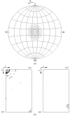
PM2 (Corticoviridae) is a dsDNA bacteriophage which contains a lipid membrane beneath its icosahedral capsid. In this respect it resembles bacteriophage PRD1 (Tectiviridae), although it is not known whether the similarity extends to the detailed molecular architecture of the virus, for instance the fold of the major coat protein P2. Structural analysis of PM2 has been initiated and virus-derived P2 has been crystallized by sitting-nanodrop vapour diffusion. Crystals of P2 have been obtained in space group P2(1)2(1)2, with two trimers in the asymmetric unit and unit-cell parameters a = 171.1, b = 78.7, c = 130.1 A. The crystals diffract to 4 A resolution at the ESRF BM14 beamline (Grenoble, France) and the orientation of the non-crystallographic threefold axes, the spatial relationship between the two trimers and the packing of the trimers within the unit cell have been determined. The trimers form tightly packed layers consistent with the crystal morphology, possibly recapitulating aspects of the arrangement of subunits in the virus.
Figures


Similar articles
-
From lows to highs: using low-resolution models to phase X-ray data.Acta Crystallogr D Biol Crystallogr. 2013 Nov;69(Pt 11):2257-65. doi: 10.1107/S0907444913022336. Epub 2013 Oct 18. Acta Crystallogr D Biol Crystallogr. 2013. PMID: 24189238 Free PMC article. Review.
-
Insights into virus evolution and membrane biogenesis from the structure of the marine lipid-containing bacteriophage PM2.Mol Cell. 2008 Sep 5;31(5):749-61. doi: 10.1016/j.molcel.2008.06.026. Mol Cell. 2008. PMID: 18775333
-
Bacteriophage PM2 has a protein capsid surrounding a spherical proteinaceous lipid core.J Virol. 2002 Aug;76(16):8169-78. doi: 10.1128/jvi.76.16.8169-8178.2002. J Virol. 2002. PMID: 12134022 Free PMC article.
-
Crystallization and preliminary X-ray analysis of receptor-binding protein P2 of bacteriophage PRD1.J Struct Biol. 2000 Aug;131(2):159-63. doi: 10.1006/jsbi.2000.4275. J Struct Biol. 2000. PMID: 11042087
-
Capsid-like arrays in crystals of chimpanzee adenovirus hexon.J Struct Biol. 2006 May;154(2):217-21. doi: 10.1016/j.jsb.2005.12.006. Epub 2006 Jan 18. J Struct Biol. 2006. PMID: 16458021 Review.
Cited by
-
Putative prophages related to lytic tailless marine dsDNA phage PM2 are widespread in the genomes of aquatic bacteria.BMC Genomics. 2007 Jul 16;8:236. doi: 10.1186/1471-2164-8-236. BMC Genomics. 2007. PMID: 17634101 Free PMC article.
-
Genetics for Pseudoalteromonas provides tools to manipulate marine bacterial virus PM2.J Bacteriol. 2008 Feb;190(4):1298-307. doi: 10.1128/JB.01639-07. Epub 2007 Dec 14. J Bacteriol. 2008. PMID: 18083813 Free PMC article.
-
From lows to highs: using low-resolution models to phase X-ray data.Acta Crystallogr D Biol Crystallogr. 2013 Nov;69(Pt 11):2257-65. doi: 10.1107/S0907444913022336. Epub 2013 Oct 18. Acta Crystallogr D Biol Crystallogr. 2013. PMID: 24189238 Free PMC article. Review.
-
Integrative Approaches to Study Virus Structures.Subcell Biochem. 2024;105:247-297. doi: 10.1007/978-3-031-65187-8_7. Subcell Biochem. 2024. PMID: 39738949 Review.
-
Genome characterization of lipid-containing marine bacteriophage PM2 by transposon insertion mutagenesis.J Virol. 2006 Sep;80(18):9270-8. doi: 10.1128/JVI.00536-06. J Virol. 2006. PMID: 16940538 Free PMC article.
References
-
- Abrescia, N. G., Cockburn, J. J., Grimes, J. M., Sutton, G. C., Diprose, J. M., Butcher, S. J., Fuller, S. D., San Martin, C., Burnett, R. M., Stuart, D. I., Bamford, D. H. & Bamford, J. K. (2004). Nature (London), 432, 68–74. - PubMed
-
- Bamford, D. H., Burnett, R. M. & Stuart, D. I. (2002). Theor. Popul. Biol.61, 461–470. - PubMed
-
- Bamford, D. H. (2003). Res. Microbiol.154, 231–236. - PubMed
-
- Benson, S. D., Bamford, J. K., Bamford, D. H. & Burnett, R. M. (2004). Mol. Cell, 16, 673–685. - PubMed
Publication types
MeSH terms
Substances
LinkOut - more resources
Full Text Sources

