. 2006 Jan 1;62(Pt 1):39-43.
doi: 10.1107/S1744309105040303. Epub 2005 Dec 16.

Crystallization of a functionally intact Hsc70 chaperone

Affiliations

Crystallization of a functionally intact Hsc70 chaperone

Jianwen Jiang et al. Acta Crystallogr Sect F Struct Biol Cryst Commun. .

Abstract

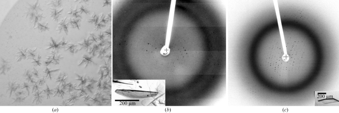
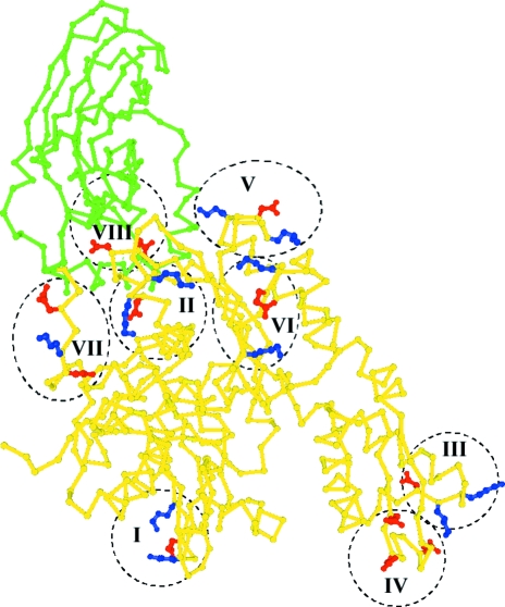
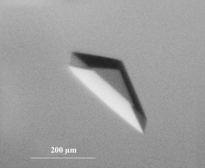
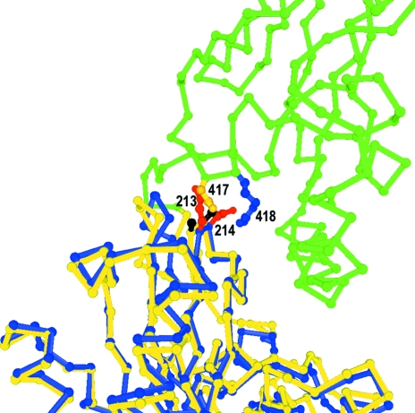
Hsp70s are essential chaperones with roles in a variety of cellular processes and representatives in all kingdoms of life. They are comprised of a nucleotide-binding domain (NBD) and a protein substrate-binding domain (SBD). Structures of isolated NBDs and SBDs have been reported but, until recently, a functionally intact Hsp70 containing both the NBD and SBD has resisted structure determination. Here, it is reported that preparation of diffraction-quality crystals of functionally intact bovine Hsc70 required (i) deletion of part of the protein to reduce oligomerization, (ii) point mutations in the interface between the SBD and NBD and (iii) use of high concentrations of the structure-stabilizing agents glycerol and trimethylamine oxide (TMAO). The introduction of point mutations in interdomain interfaces and the use of the potent structure stabilizer TMAO may be generally useful in crystallization of multidomain proteins that exhibit interdomain motions.


Figures

Figure 1
Figure 1
Deletion of the 10 kDa C-terminal domain of Hsc70 reduces oligomerization. Lanes 1 and 2: SDS–PAGE of Hsc70ΔCterm and full-length Hsc70, respectively. Lanes 3 and 4: native PAGE of Hsc70ΔCterm and full-length Hsc70, respectively.
Figure 2
Figure 2
(a) Hsc70ΔCterm crystals obtained in initial screens (microbatch under oil: 1 µl of 20 mg ml−1 protein in 10 mM Tris pH 8.0, 1 mM EDTA, 1 mM DTT, 50% glycerol plus 1 µl 0.2 M sodium acetate, 0.1 M Tris 8.5, 30% PEG 4000). (b) Optimized crystals [inset; grown as in (a) but with 0.5 M KI added] and diffraction pattern. (c) Further optimization of crystals [inset; grown as in (b) but the protein was further purified by cation-exchange and gel-exclusion chromatography] and a representative diffraction pattern (P212121; a = 78.3, b = 95.2, c = 178.5 Å).
Figure 3
Figure 3
Crystal structure of bovine Hsc70ΔCterm with NBD in yellow and SBD in green and residues mutated to alanines highlighted. I, K108/E110/K112; II, D186/K187/K188; III, K250/K251/D252; IV, E283/D285/E289; V, K325/D327/K328; VI, K357/E358/K361; VII, D383/K384/E386; VIII, E213/D214.
Figure 4
Figure 4
Native PAGE of wild-type (WT) and mutant bHsc70ΔCterms. Above each lane the type of residues mutated and the net effect on the protein’s charge are given (i.e. in lane 1 DKK to AAA changes one negative and two positive side chains to neutral, leading to a net change of −1). Changes in electrophoretic migration are consistent with the expected net changes in charge.
Figure 5
Figure 5
Crystal of Hsc70ΔCtermE213A/D214A mutant (macrobatch under oil at 289 K: 5 µl of 13 mg ml−1 protein in 10 mM Tris pH 8.0, 50% glycerol, 1 mM EDTA, 1 mM DTT plus 5 µl of 4 M TMAO plus 10 µl of 18% PEG 8000).
Figure 6
Figure 6
Location of E213A/D214A mutations at the interdomain interface. The interface between the SBD (green) and NBD (yellow) from the Hsc70ΔCtermE213A/D214A structure is shown with the A213/A214 mutant side chains in black and Asn417 and Arg418 in yellow and blue, respectively. Superimposed on this structure is the Cα trace for the isolated bovine Hsc70 ATPase domain (in blue) with the E213/D214 side chains in red.

References

    1. Auton, M. & Bolen, D. W. (2005). Proc. Natl Acad. Sci. USA, 102, 15065–15068. - PMC - PubMed
    1. Banecki, B., Zylicz, M., Bertoli, E. & Tanfani, F. (1992). J. Biol. Chem.267, 25051–25058. - PubMed
    1. Baskakov, I. V. & Bolen, D. W. (1998). J. Biol. Chem.273, 4831–4834. - PubMed
    1. Baskakov, I. V., Kumar, R., Srinivasan, G., Ji, Y.-S., Bolen, D. W. & Thompson, E. B. (1999). J. Biol. Chem.274, 10693–10696. - PubMed
    1. Boice, J. A. & Hightower, L. E. (1997). J. Biol Chem.272, 24825–24831. - PubMed

Publication types

Associated data