. 2006 Apr 5;25(7):1559-68.
doi: 10.1038/sj.emboj.7601034. Epub 2006 Mar 2.

Alternative intermolecular contacts underlie the rotavirus VP5* two- to three-fold rearrangement

Affiliations

Alternative intermolecular contacts underlie the rotavirus VP5* two- to three-fold rearrangement

Joshua D Yoder et al. EMBO J. .

Abstract

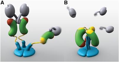
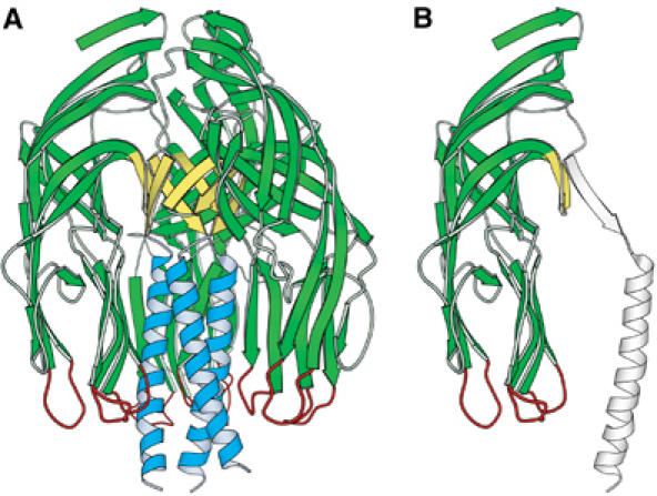
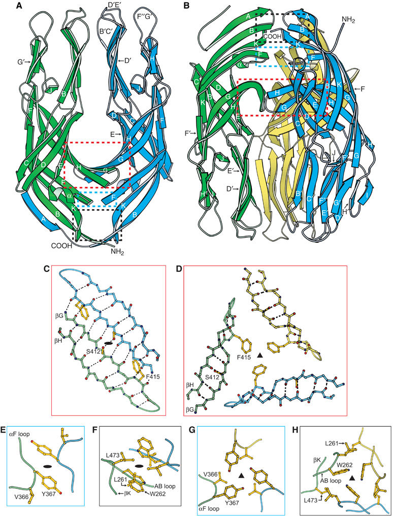
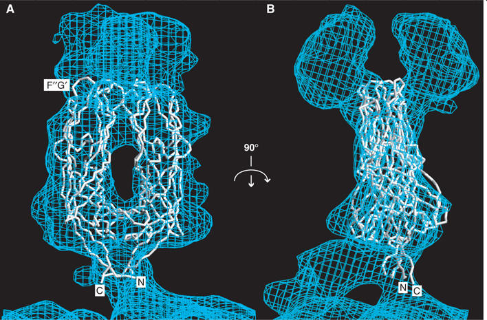
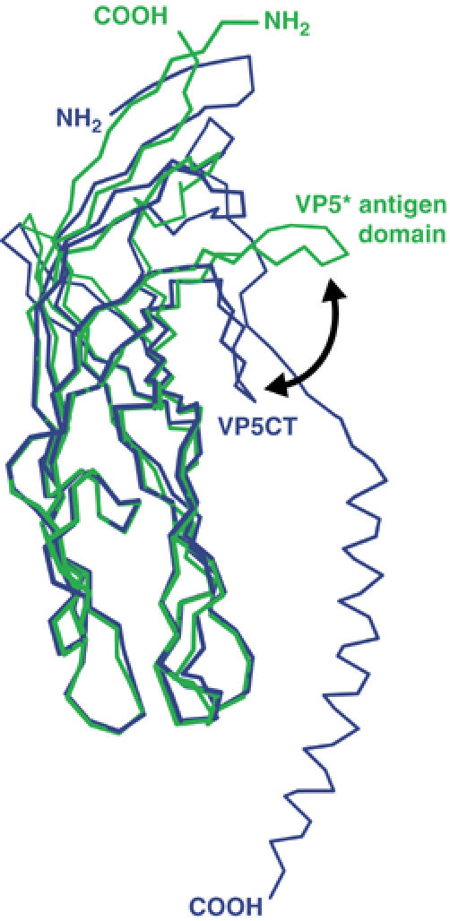
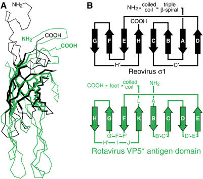
The spike protein VP4 is a key component of the membrane penetration apparatus of rotavirus, a nonenveloped virus that causes childhood gastroenteritis. Trypsin cleavage of VP4 produces a fragment, VP5*, with a potential membrane interaction region, and primes rotavirus for cell entry. During entry, the part of VP5* that protrudes from the virus folds back on itself and reorganizes from a local dimer to a trimer. Here, we report that a globular domain of VP5*, the VP5* antigen domain, is an autonomously folding unit that alternatively forms well-ordered dimers and trimers. Because the domain contains heterotypic neutralizing epitopes and is soluble when expressed directly, it is a promising potential subunit vaccine component. X-ray crystal structures show that the dimer resembles the spike body on trypsin-primed virions, and the trimer resembles the folded-back form of the spike. The same structural elements pack differently to form key intermolecular contacts in both oligomers. The intrinsic molecular property of alternatively forming dimers and trimers facilitates the VP5* reorganization, which is thought to mediate membrane penetration during cell entry.


Figures

Figure 1
Figure 1
Models of two VP4 conformations. (A) The primed state. Two rigid subunits form the spike visible in electron cryomicroscopy image reconstructions of trypsin-primed virions. A third subunit is flexible. VP8* is gray, with an N-terminal tether and a globular head creased by the sialoside-binding site. The VP5* antigen domain is green bean-shape, with a red membrane interaction region and a yellow GH loop. An additional β-strand C-terminal to the antigen domain is also yellow. The spike body includes the VP5* antigen domain, part of the VP8* tether, and the GH loop. The foot is blue, as is a protruding region that rearranges into the coiled-coil. (B) The putative post-membrane penetration state. VP8* has dissociated; the yellow parts of each subunit have joined in a β-annulus; the α-helical triple coiled-coil has zipped up; and the VP5* antigen domain has folded back. The models were produced by Digizyme, Inc.
Figure 2
Figure 2
VP5CT and the VP5* antigen domain. (A) Ribbon diagram of the VP5CT trimer, colored to match Figure 1. VP5CT does not include the foot region. (B) Ribbon diagram of a single VP5CT subunit. The part that forms the VP5* antigen domain is green, yellow, and red; the remainder is drawn in outline.
Figure 3
Figure 3
Crystal structures of the VP5* antigen domain dimer and trimer. (A) Ribbon diagram of the dimer. The orientation matches the model of the rigid spikes on the virion in Figure 1A. The red, blue, and black boxes show the regions detailed in panels (C), (E), and (F), respectively. The termini of the green subunit are indicated. Secondary structural elements, including the three hydrophobic loops of the blue subunit are labeled. (B) Ribbon diagram of the trimer. The orientation matches the model of the rearranged trimer in Figure 1B. The red, blue, and black boxes show the regions detailed in panels (D), (G), and (H), respectively. The termini of the blue subunit are indicated. (C, E, F) Atomic details of the key intersubunit contacts of the dimer. Black ovals indicate the approximate two-fold axis. In panel (F), the L261 side chain is below the W262 side chain. (D, G, H) Atomic details of the key intersubunit contacts of the trimer. Black triangles indicate the three-fold axis. In panel (H), the W262 rings are seen on-edge with the five-atom pyrrole ring closest to the viewer. Panels (C)–(H) are drawn as if looking down from the tops of the ribbon diagrams in panels (A) and (B). Panels (C) and (D), panels (E) and (G), and panels (F) and (H) are pairs, showing alternative packing of the same residues. The depicted side chains are discussed in the text. Dashed black lines indicate hydrogen bonds.
Figure 4
Figure 4
The VP5* antigen domain dimer fit to the molecular envelope of the primed spike. The molecular envelope of an approximately 12 Å resolution electron cryomicroscopy image reconstruction of a VP4 spike on a trypsin-primed SA11-4F rotavirus virion is contoured at 0.5 σ. (A) Depicted from the perspective of Figures 1A and 3A. The Cα trace includes residues T259-N477 of one subunit (on the left) and residues T259-S476 of the second subunit (on the right). The termini and the F″G′ loop of one subunit are indicated. (B) Rotated 90° about a vertical axis from panel (A).
Figure 5
Figure 5
Superposition of the VP5* antigen domain and VP5CT. Residues S260-S476 of a VP5* antigen domain subunit in the dimer conformation are drawn as a green Cα trace. Residues I254-L522 of a VP5CT subunit are drawn as a blue Cα trace. The black arrow indicates the movement of the GH loop between the two states.
Figure 6
Figure 6
Comparison of the rotavirus VP5* antigen domain and the reovirus σ1 knob. (A) Superposition of Cα traces. Residues Y298 to T455 of reovirus σ1 (black; PDB identification code 1KKE) are superimposed on residues I254-L473 of a subunit of the rotavirus VP5* antigen domain trimer (green). The residues used to calculate the superposition matrix are drawn with thick lines. (B) Folding diagram of the β-barrel of reovirus σ1 and the β-sandwich of rotavirus VP5*. Arrows depict β-strands.

References

    1. Arias CF, Romero P, Alvarez V, Lopez S (1996) Trypsin activation pathway of rotavirus infectivity. J Virol 70: 5832–5839 - PMC - PubMed
    1. Brunger AT, Adams PD, Clore GM, DeLano WL, Gros P, Grosse-Kunstleve RW, Jiang JS, Kuszewski J, Nilges M, Pannu NS, Read RJ, Rice LM, Simonson T, Warren GL (1998) Crystallography & NMR system: a new software suite for macromolecular structure determination. Acta Crystallogr D 54: 905–921 - PubMed
    1. Chappell JD, Prota AE, Dermody TS, Stehle T (2002) Crystal structure of reovirus attachment protein sigma1 reveals evolutionary relationship to adenovirus fiber. EMBO J 21: 1–11 - PMC - PubMed
    1. Ciarlet M, Ludert JE, Iturriza-Gomara M, Liprandi F, Gray JJ, Desselberger U, Estes MK (2002) The initial interaction of rotavirus strains with N-acetyl-neuraminic (sialic) acid residues on the cell surface correlates with VP4 genotype, not species of origin. J. Virol 76: 4087–4095 - PMC - PubMed
    1. Collaborative Computational Project (1994) The CCP4 Suite: programs for protein crystallography. Acta Crystallogr D 50: 760–763 - PubMed

Publication types

LinkOut - more resources