. 2006 Mar;116(3):663-74.
doi: 10.1172/JCI26027.

Tbx18 regulates the development of the ureteral mesenchyme

Affiliations

Tbx18 regulates the development of the ureteral mesenchyme

Rannar Airik et al. J Clin Invest. 2006 Mar.

Abstract

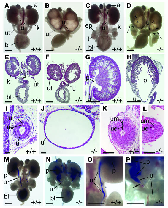
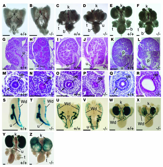
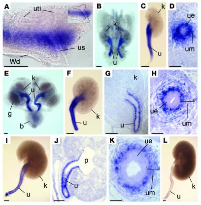
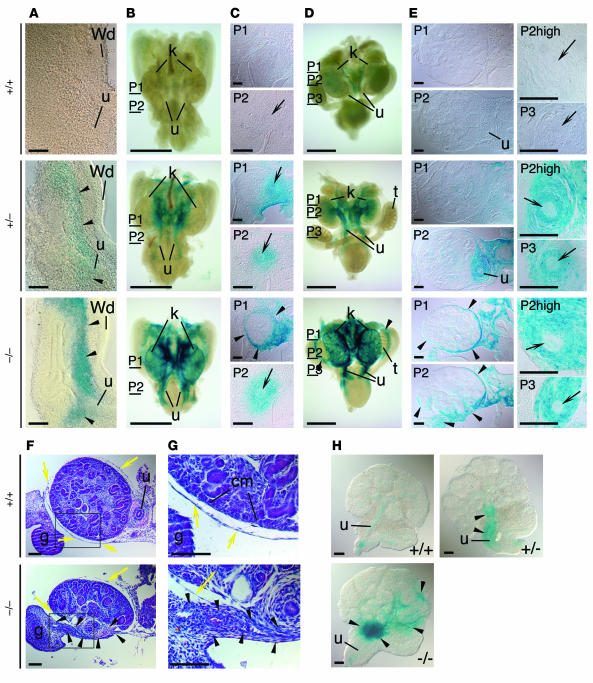
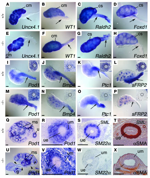
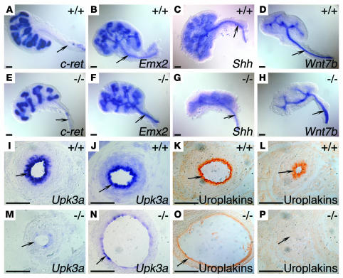
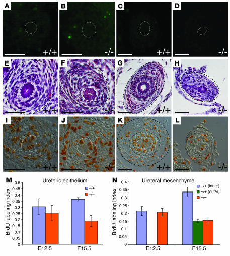
Congenital malformations of the urinary tract are a major cause of renal failure in children and young adults. They are often caused by physical obstruction or by functional impairment of the peristaltic machinery of the ureter. The underlying molecular and cellular defects are, however, poorly understood. Here we present the phenotypic characterization of a new mouse model for congenital ureter malformation that revealed the molecular pathway important for the formation of the functional mesenchymal coating of the ureter. The gene encoding the T-box transcription factor Tbx18 was expressed in undifferentiated mesenchymal cells surrounding the distal ureter stalk. In Tbx18-/- mice, prospective ureteral mesenchymal cells largely dislocalized to the surface of the kidneys. The remaining ureteral mesenchymal cells showed reduced proliferation and failed to differentiate into smooth muscles, but instead became fibrous and ligamentous tissue. Absence of ureteral smooth muscles resulted in a short hydroureter and hydronephrosis at birth. Our analysis also showed that the ureteral mesenchyme derives from a distinct cell population that is separated early in kidney development from that of other mesenchymal cells of the renal system.


Figures

Figure 1
Figure 1
Kidney and urogenital tract anomalies in Tbx18–/– embryos at E18.5. (AD) Morphology of whole urogenital systems of female (A and B) and male embryos (C and D). Arrowheads in C and D indicate blood vesels. Arrows in D indicate ligaments connecting the testes to the kidneys. (EL) Hematoxylin and eosin stainings of sagittal sections of whole urogenital systems (E and F) and kidneys (G and H) and of transverse sections of the ureter (IL). (I and K) The ureter epithelium and its surrounding mesenchyme were multilayered in the wild-type embryo at the kidney level (I) and more distally (K). (J and L) In the Tbx18–/– ureter, a single-layered epithelium was surrounded by a thin layer of mesenchymal cells at proximal (J) and distal (L) levels. (MP) Physical obstruction of the Tbx18–/– ureter as revealed by ink injection experiments. (O and P) Higher-magnification views of the ureteropelvic region shown in M and N. Arrow in P indicates obstruction in the ureter. a, adrenal; bl, bladder; ep, epididymis; k, kidney; p, pelvis; t, testis; u, ureter; ue, ureteric epithelium; um, ureteral mesenchyme; ut, uterus. Scale bars: 1,000 μm (AH and MP), 100 μm (IL).
Figure 2
Figure 2
Early onset of kidney and ureter anomalies in Tbx18–/– embryos. Morphology of whole urogenital systems (AF), hematoxylin and eosin stainings of sagittal sections of kidneys (GL), and transverse sections of ureters (MR) at E12.5 (A, B, G, H, M, and N), E14.5 (C, D, I, J, O, and P), and E16.5 (E, F, K, L, Q, and R). Arrows in M, O, and Q indicate an inner ring of condensed mesenchymal cells around the ureteric epithelium that was missing in the mutant (N, P, and R). (SZ) β-Galactosidase activity staining of kidneys at E11.5 (S and T) and of whole urogenital systems at E12.5 (U and V), E14.5 (W and X), and E18.5 (Y and Z) of a Pax2:LacZ reporter gene driving expression exclusively into the epithelium of Wolffian duct (Wd) and its derivative, the ureter. Hydroureter and hydronephrosis occurred after onset of urine formation at E16.5 (F, L, and R) and were clearly developed by E18.5 (Z). Scale bars: 500 μm (AF, IL, and SZ), 50 μm (G, H, and MR).
Figure 3
Figure 3
Tbx18 expression is confined to the developing ureter. In situ hybridization analysis of Tbx18 expression in kidneys at E11.5 (A), E12.5 (C), E14.5 (F), E16.5 (I), and E18.5 (L), in whole urogenital systems at E12.5 (B) and E14.5 (E), on transverse sections of ureters at E12.5 (D), E14.5 (H), and E16.5 (K), and on longitudinal sections of kidneys with attached ureters of wild-type embryos at E14.5 (G) and E16.5 (J). The basal surface of the ureteric epithelium is outlined (A, inset). Tbx18 expression was confined to prospective and definitive periureteral mesenchymal cells and excluded from the ureteric epithelium. Expression appeared higher in the inner layer of ureteral mesenchymal cells than in the outer ring (H and K). Expression was downregulated with differentiation of mesenchymal cells into SMCs after E14.5. g, gonad; uti, ureter tip; us, ureter stalk. Scale bars: 200 μm (AC, EG, I, J, and L), 100 μm (D, H, and K).
Figure 4
Figure 4
Mislocalization of Tbx18:LacZ expression in Tbx18–/– urogenital systems. (AE) β-Galactosidase activity staining of a LacZ reporter gene in the Tbx18 locus was analyzed in kidneys at E11.5 (A), in urogenital systems at E12.5 (B) and E14.5 (D), and in transverse sections of E12.5 (C) and E14.5 kidneys (E). P1 and P2 in C indicate sections from the planes shown in B. P1, P2, and P3 in E indicate section planes shown in D. P2high is a higher magnification of P2 in the region of the ureter. Black arrows indicate the ureteric epithelium. (F and G) Hematoxylin and eosin stainings of transverse sections of E14.5 kidneys. Yellow arrows indicate the renal capsule. The boxed regions in F are magnified in G. (H) β-Galactosidase activity staining of 2-day-old cultures of metanephric rudiments that were wild type as well as heterozygous and homozygous for the mutant Tbx18LacZ allele. Slight blue staining of the ureteric epithelium in wild-type mice was due to endogenous β-galactosidase activity. Heterozygotes and homozygotes in A, B, and D were Tbx18LacZ/+ and Tbx18LacZ/Tbx18LacZ, respectively, whereas embryos used for C and E were normalized for the LacZ allele (i.e., Tbx18LacZ/+ and Tbx18/Tbx18LacZ, respectively). cm, condensing (metanephric) mesenchyme. Arrowheads in A and CH mark localization of Tbx18-positive prospective and definitive ureteral mesenchymal cells in kidneys, urogenital systems, and kidney cultures at the indicated stages. Scale bars: 100 μm (A, C, and EH), 1,000 μm (B and D).
Figure 5
Figure 5
Molecular characterization of Tbx18–/– ureteral mesenchyme. Whole-mount in situ hybridization analysis of mesenchymal marker genes in E12.5 kidneys and ureters (AP). Arrows indicate ureteral mesenchyme. Insets in (IP) show in situ hybridization analysis on transverse ureter sections. Ptc1, Patched1. The ureteric epithelium is shown by a black circle. In situ hybridization analysis of Pod1 expression on transverse sections of E14.5 kidneys (Q and U) and ureters (R and V) and of SM22α expression on transverse sections of E16.5 ureters (S and W). Arrowheads in U indicate ectopic fibrous tissue connecting gonad and kidney. (T and X) Immunohistochemistry for α-SMA on transverse sections of E18.5 ureters. cs, cortical stroma; ms, medullary stroma; SML, smooth muscle layer. Scale bars: 100 μm, 120 μm (insets).
Figure 6
Figure 6
Molecular characterization of the Tbx18–/– ureteric epithelium. (AH) Whole-mount in situ hybridization analysis at E12.5. (I, J, M, and N) In situ hybridization analysis on transverse ureter sections at E16.5 (I and M) and E18.5 (J and N) and (K, L, O, and P) immunohistochemistry on transverse sections of E18.5 ureters at a proximal position (K and O) and more distally (L and P) of Tbx18–/– embryos and control wild-type littermates. Arrows indicate ureteric epithelium. Scale bars: 100 μm.
Figure 7
Figure 7
Apoptosis and proliferation in Tbx18–/– ureters. (AD) TUNEL assay on transverse sections of E12.5 (A and B) and E15.5 (C and D) ureters did not reveal a difference in apoptosis between ureteric epithelium (within dashed outline) and the ureteral mesenchyme (outside dashed outline) of Tbx18 mutant embryos (B and D) and wild-type littermates (A and C). (EH) Hematoxylin and eosin stainings of transverse sections of wild-type and Tbx18–/– ureters at E12.5 (E and F) and E15.5 (G and H). (IL) Analysis of cell proliferation in transverse sections of the ureter of wild-type (I and K) and Tbx18 mutant embryos (J and L) at E12.5 (I and J) and E15.5 (K and L) by the BrdU incorporation assay. In EL, dashed white outlines show the ureteric epithelium and dashed black outlines the inner and outer layers of the ureteral mesenchyme as determined by the border of highly condensed mesenchymal cells and tangentially oriented loose mesenchymal cells, respectively. (M and N) Quantification of cell proliferation by the BrdU labeling index at E12.5 and E15.5 in the analyzed ureteral mesenchyme (N) and in the ureteric epithelium (M). In E12.5 epithelium, P = 0.30, wild-type (0.31 ± 0.07) versus mutant (0.25 ± 0.06). In E12.5 mesenchyme, P = 0.71, wild-type (0.21 ± 0.03) versus mutant (0.21 ± 0.02). In E15.5 epithelium, P < 0.0003, wild-type (0.37 ± 0.01) versus mutant (0.19 ± 0.04). In E15.5 mesenchyme, P < 0.00004, wild-type inner ring (0.34 ± 0.03) versus mutant (0.16 ± 0.02); P = 0.77, wild-type outer ring (0.15 ± 0.01) versus mutant (0.16 ± 0.02). Scale bars: 50 μm.

Comment in

Similar articles

Cited by

References

    1. Kohaut EC, Tejani A. The 1994 annual report of the North American Pediatric Renal Transplant Cooperative Study. Pediatr. Nephrol. 1996;10:422–434. - PubMed
    1. Woolf AS, Winyard PJ. Molecular mechanisms of human embryogenesis: developmental pathogenesis of renal tract malformations. Pediatr. Dev. Pathol. 2002;5:108–129. - PubMed
    1. Miyazaki Y, Ichikawa I. Ontogeny of congenital anomalies of the kidney and urinary tract, CAKUT. Pediatr. Int. 2003;45:598–604. - PubMed
    1. Chevalier RL. Perinatal obstructive nephropathy. Semin. Perinatol. 2004;28:124–131. - PubMed
    1. Tanagho, E.A. 1981. Development of the ureter. In The ureter. H. Bergman, editor. Springer–Verlag. New York, New York, USA. 1–12.

Publication types

MeSH terms

Substances