. 2006 Mar;116(3):675-82.
doi: 10.1172/JCI27374.

Local InsP3-dependent perinuclear Ca2+ signaling in cardiac myocyte excitation-transcription coupling

Affiliations

Local InsP3-dependent perinuclear Ca2+ signaling in cardiac myocyte excitation-transcription coupling

Xu Wu et al. J Clin Invest. 2006 Mar.

Abstract

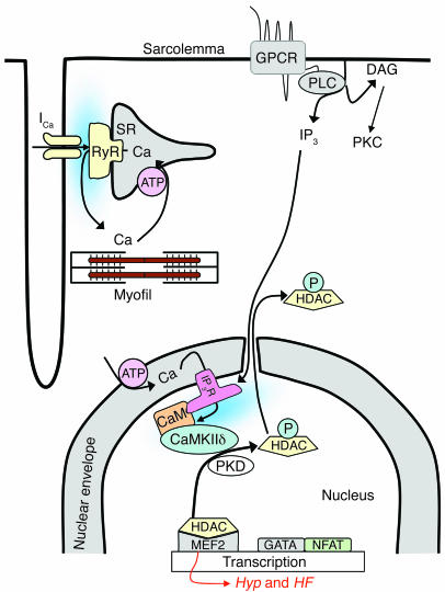
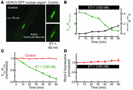
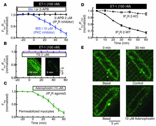
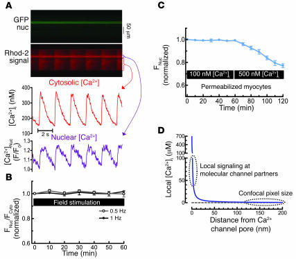
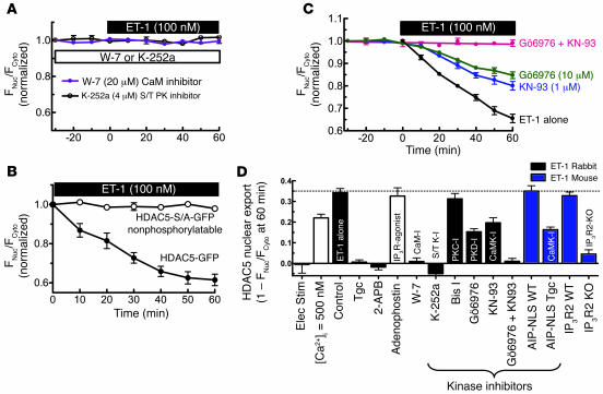
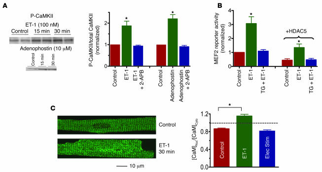
Previous work showed that calmodulin (CaM) and Ca2+-CaM-dependent protein kinase II (CaMKII) are somehow involved in cardiac hypertrophic signaling, that inositol 1,4,5-trisphosphate receptors (InsP3Rs) in ventricular myocytes are mainly in the nuclear envelope, where they associate with CaMKII, and that class II histone deacetylases (e.g., HDAC5) suppress hypertrophic gene transcription. Furthermore, HDAC phosphorylation in response to neurohumoral stimuli that induce hypertrophy, such as endothelin-1 (ET-1), activates HDAC nuclear export, thereby regulating cardiac myocyte transcription. Here we demonstrate a detailed mechanistic convergence of these 3 issues in adult ventricular myocytes. We show that ET-1, which activates plasmalemmal G protein-coupled receptors and InsP3 production, elicits local nuclear envelope Ca2+ release via InsP3R. This local Ca2+ release activates nuclear CaMKII, which triggers HDAC5 phosphorylation and nuclear export (derepressing transcription). Remarkably, this Ca2+-dependent pathway cannot be activated by the global Ca2+ transients that cause contraction at each heartbeat. This novel local Ca2+ signaling in excitation-transcription coupling is analogous to but separate (and insulated) from that involved in excitation-contraction coupling. Thus, myocytes can distinguish simultaneous local and global Ca2+ signals involved in contractile activation from those targeting gene expression.


Figures

Figure 1
Figure 1
Working hypothesis. ET-1 induces HDAC5 nuclear export via a pathway involving InsP3 (IP3) and InsP3R (IP3R) with associated CaM and CaMKII, leading to HDAC5 phosphorylation and nuclear export, thereby derepressing the transcription factor MEF2 (see text for additional details). Hyp, hypertrophy.
Figure 2
Figure 2
ET-1 induces nuclear export of HDAC5. (A) ET-1 (100 nM) was applied for 60 minutes to quiescent rabbit ventricular myocyte expressing the fusion protein HDAC5-GFP. (B) Individual traces of nuclear and cytosolic fluorescence (FNuc/FCyto) for a single myocyte, where each trace is normalized to the Fcyto before ET-1 exposure (Fcyto-initial). (C) HDAC5 nuclear export was analyzed as decrease of FNuc/FCyto, normalized to the initial ratio (6.3 ± 0.4; n = 12). The control group was treated the same, except without ET-1 application (n = 10). (D) [Ca2+]i measured by Rhod-2 fluorescence upon exposure to 100 nM ET-1 (n = 7). Long-term exposure to ET-1 induced further HDAC5 export, such that after 24 hours the nucleus was virtually depleted of HDAC5 (not shown), analogous to effects in cultured neonatal rat myocytes (31).
Figure 3
Figure 3
ET-1–induced HDAC5 nuclear export is dependent on InsP3R and Ca2+ in InsP3-sensitive stores. (A) HDAC5-GFP–expressing myocytes were pretreated with 2 μM 2-APB (n = 6) or 10 μM Bis I (n = 5) for 30 minutes, followed by application of 100 nM ET-1. (B) SR and nuclear envelope Ca2+ stores were depleted by preincubation with SR/ER Ca2+-ATPase (SERCA) inhibitor TG 1 μM (n = 7) for 10 minutes. Insets are the Fluo-5N images, showing that this TG treatment depleted nuclear envelope Ca2+ stores. (C) In permeabilized HDAC5-GFP expressing myocytes, 10 μM adenophostin was applied after 30 minutes at 100 nM [Ca2+]i (n = 8). HDAC5 nuclear export was measured as the decrease of nuclear fluorescence. In the permeabilized cell, cytosolic concentration is irrelevant as HDAC5-GFP can readily diffuse to the bath. (D) ET-1–induced HDAC5 nuclear export assessed in mouse ventricular myocytes that lack InsP3R2 (InsP3R 2 KO) or WT littermates. (E) Fluo-5N–loaded myocytes were permeabilized and treated without or with 10 μM adenophostin for 30 minutes. Nuclear envelope Ca2+ release was indicated by decreased Fluo-5N fluorescence.
Figure 4
Figure 4
HDAC5 nuclear export is Ca2+ dependent but not activated by global Ca2+ transients. (A) Adenoviral HDAC5-GFP–infected myocytes were also loaded with Rhod-2 to measure [Ca2+]i. Rhod-2 is more concentrated in the nucleus (nuc), complicating Ca2+ transient calibration there. (B) HDAC5 remained nuclear when myocytes were field stimulated at 0.5 Hz (n = 5) or 1 Hz (n = 7) for 60 minutes or even at 2 Hz (not shown). (C) In permeabilized myocytes, internal [Ca2+] was increased from 100 nM to 500 nM (at 60 minutes) (n = 7). 2,3 butanedione monoxime (5 mM) was used to prevent contraction. (D) Expected local [Ca2+] gradient around the mouth of an InsP3R Ca2+ channel: [Ca2+]i = [Ca2+]Init + q/(2πDr) × erfc{r/(2√Dr)}, where q = single channel current (0.1 pA), D = diffusion coefficient (600 μm2/s), [Ca2+]Init = 100 nM, erfc = complementary error function, and r = radial distance from the channel mouth for hemispheric diffusion. This is the steady state, achieved in approximately 10 μs without buffering and much less than 1 ms when local Ca2+ buffering is included (26).
Figure 5
Figure 5
Role of CaM and protein kinase in ET-1–induced HDAC5 nuclear export. (A) Myocytes were preincubated for 30 minutes with 4 μM K-252a (n = 8) or 20 μM W-7 (n = 7) followed by ET-1 (100 nM) application. (B) ET-1–induced nuclear export of HDAC5-GFP versus mutant nonphosphorylatable HDAC5 (HDAC5-S/A-GFP; n = 4). (C) Adv-GFP-HDAC5–infected myocytes were pretreated with 1 μM KN-93 (n = 6) or 10 μM Gö6976 (n = 8), separately or simultaneously for 20–30 minutes followed by ET-1 application. (D) Summary of HDAC5 nuclear export under different conditions. Elec stim, electrical stimulation; S/T PK, serine/threonine protein kinase; Tgc, transgenic.
Figure 6
Figure 6
CaMKII autophosphorylation, MEF2 activity, and CaM translocation. (A) Adult rabbit myocytes were treated with 100 nM ET-1 or 10 μM adenophostin as in Figures 2B and 3C, with cells quickly frozen for CaMKII autophosphorylation measurement by Western blot (n = 3 for each group). (B) Adenoviral MEF2-luciferase reporter was expressed in rabbit ventricular myocyte (± Adv-HDAC5-GFP) and challenged for 1 hour with 100 nM ET-1 (± pretreatment for 10 minutes with 1 μM TG). Luciferase activity was normalized to control (without HDAC5-GFP). (C) Rabbit myocytes ± ET-1, with CaM movement detected by immunocytochemistry during rest (control), exposure to 100 nM ET-1, or electrical stimulation at 1 Hz (n = 8 for each group). *P < 0.05.

Comment in

References

    1. Frey N, McKinsey TA, Olson EN. Decoding calcium signals involved in cardiac growth and function. Nat. Med. 2000;6:1221–1227. - PubMed
    1. Crabtree GR, Olson EN. NFAT signaling: choreographing the social lives of cells. Cell. 2002;109:S67–S79. - PubMed
    1. Zhu W, et al. Ca2+/calmodulin-dependent kinase II and calcineurin play critical roles in endothelin-1-induced cardiomyocyte hypertrophy. J. Biol. Chem. 2000;275:15239–15245. - PubMed
    1. Gruver CL, DeMayo F, Goldstein MA, Means AR. Targeted developmental overexpression of calmodulin induces proliferative and hypertrophic growth of cardiomyocytes in transgenic mice. Endocrinology. 1993;133:376–388. - PubMed
    1. Colomer JM, Means AR. Chronic elevation of calmodulin in the ventricles of transgenic mice increases the autonomous activity of calmodulin-dependent protein kinase II, which regulates atrial natriuretic factor gene expression. Mol. Endocrinol. 2000;14:1125–1136. - PubMed

Publication types

MeSH terms

Substances