. 2006 Mar;116(3):760-8.
doi: 10.1172/JCI25303.

Loss of constitutive activity of the growth hormone secretagogue receptor in familial short stature

Affiliations

Loss of constitutive activity of the growth hormone secretagogue receptor in familial short stature

Jacques Pantel et al. J Clin Invest. 2006 Mar.

Abstract

The growth hormone (GH) secretagogue receptor (GHSR) was cloned as the target of a family of synthetic molecules endowed with GH release properties. As shown recently through in vitro means, this receptor displays a constitutive activity whose clinical relevance is unknown. Although pharmacological studies have demonstrated that its endogenous ligand--ghrelin--stimulates, through the GHSR, GH secretion and appetite, the physiological importance of the GHSR-dependent pathways remains an open question that gives rise to much controversy. We report the identification of a GHSR missense mutation that segregates with short stature within 2 unrelated families. This mutation, which results in decreased cell-surface expression of the receptor, selectively impairs the constitutive activity of the GHSR, while preserving its ability to respond to ghrelin. This first description, to our knowledge, of a functionally significant GHSR mutation, which unveils the critical importance of the GHSR-associated constitutive activity, discloses an unusual pathogenic mechanism of growth failure in humans.


Figures

Figure 1
Figure 1
Identification of a GHSR mutation. (A) Electrophoregram spanning the GHSR mutation site from a proband presenting with ISS and a control. The sequence variation, present in the homozygous state, is a C-to-A transversion located within the first GHSR exon (c.611C→A). (B) Location of the predicted A204E mutation in the second extracellular loop of the GHSR1a, a 7-transmembrane G-coupled receptor. (C) Conservation of the GHSR1a amino acid sequence among species within the region bracketing the mutation site between the fourth (TM4) and the fifth (TM5) transmembrane domains; A204E is shown by an arrow.
Figure 2
Figure 2
Inheritance of the A204E GHSR mutation in families 1 and 2. (A) Family 1. (B) Family 2. Circles and squares denote female and male family members, respectively. The SD to mean height for age is given below each symbol; height values are before GH treatment. Black symbols denote a short stature. The probands are indicated by arrows. The segregation of the GHSR A204E allele within both families was carried out by means of a specific restriction fragment length polymorphism (the A204E mutation creates an MnlI site).
Figure 3
Figure 3
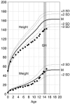
Height (cm) and weight (kg) curves of the proband (family 1, patient II2). The GH treatment is shown by the gray area.
Figure 4
Figure 4
Expression of the A204E mutant GHSR1a at the cellular level. (A) Specific binding of 125I-ghrelin to HEK293 cells transiently transfected with empty vector (mock), or HA-GHSR-WT (WT) or HA-GHSR-A204E (A204E), as determined on whole cells. Transfected cells were incubated over 4 hours at 4°C with 125I-ghrelin (30 pM). The specific binding, which represents the difference between total binding and nonspecific binding, is expressed as a percentage of the binding associated with the WT GHSR1a receptor. A representative experiment of 3 independent experiments (each performed in triplicate) is shown. (B) Displacement curves for 125I-ghrelin binding to whole HEK293 cells stably expressing the A204E mutant or the WT GHSR1a. The binding of 30 pM of 125I-ghrelin was displaced by increasing concentrations of cold ghrelin. A representative experiment of 3 independent experiments (each performed in triplicate) is shown. (C) Immunolocalization of HA-tagged WT and A204E mutant receptors transiently expressed in COS-7 cells (×40). The immunostaining of HA-tagged GHSR1a receptors was performed by means of an anti-HA monoclonal primary antibody incubated in the absence or in the presence of a permeabilizing reagent in order to visualize the receptors located at the cell surface or at both the cell surface and the intracellular level, respectively. Anti-HA mAb labeling was revealed by an Alexa Fluor 568 goat anti-mouse secondary antibody. Merge pictures show the anti-HA Alexa Fluor 568 staining (red) together with the staining of nuclei with DAPI (blue).
Figure 5
Figure 5
A204E GHSR1a agonist-independent signal transduction via the CRE pathway in transiently transfected HEK293 cells. (A) Constitutive activity of GHSR1a as a function of its own cell-surface expression. The constitutive activity associated with increasing concentrations of WT or A204E GHSR1a vectors was assessed in cotransfection experiments with an HA-tagged GHSR1a plasmid (HA-GHSR-WT or HA-GHSR-A204E) and a CRE-containing reporter plasmid (pPOU1F1-Luc). The corresponding luminometric (RLU) signal was normalized to protein concentration (RLU/μg proteins). The HA-GHSR1a cell-surface expression detected by means of an anti-HA epitope antibody (ELISA) is expressed in OD units. Signals associated with cells transfected with the mock plasmid have been subtracted, so that plotted signals represent specific RLU. Values are the mean ± SD of 1 representative experiment performed in 4 replicates among 3 independent experiments. (B) Evaluation of a putative dominant-negative effect of the A204E mutant on the WT GHSR1a. Constitutive activity in cells expressing the GHSR1a WT, the GHSR1a A204E mutant, or both isoforms is expressed as a percentage of the basal activity of the WT GHSR1a receptor. Values are the mean ± SD of 3 independent experiments, each performed in triplicate.
Figure 6
Figure 6
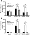
Ligand-mediated signal transduction of the A204E GHSR1a in transiently transfected HEK293 cells. Cells expressing WT GHSR1a, GHSR1a A204E, or mock-transfected cells were incubated either in the absence or in the presence of an agonist (ghrelin at 10–6 M), or of an inverse agonist ([D-Arg1, D-Phe5, D-Trp7,9, Leu11]-substance P, or SPA, at 10–6 M). The transcriptional activity in each condition (RLU/μg proteins) is expressed as a percentage of the basal activity of the WT GHSR1a receptor. Values are the mean ± SD of 1 representative experiment performed in triplicate among 6 independent experiments. *P < 0.05 and **P < 0.01 (ligand-induced signal versus basal level). (A) CRE-mediated transcriptional activity. Cells were cotransfected with 100 ng of each expression plasmid and 250 ng of the pPOU1F1-Luc reporter plasmid. (B) SRE-mediated transcriptional activity. Cells were cotransfected with 10 ng of each expression plasmid and 500 ng of the pSRE-Luc reporter plasmid.
Figure 7
Figure 7
Calcium response to ghrelin of the WT and A204E GHSR1a receptors stably expressed in HEK293 cells. The HEK293 clones expressing the WT or the mutant receptor were selected on their similar GHSR1a expression pattern. GHSR1a-expressing cells and untransfected control cells were stimulated with increasing concentrations of ghrelin. The intracellular Ca2+ release was monitored by means of the Fluo-4 AM fluorescent probe. Each signal, which represents the mean of triplicates, is expressed as the percentage of total calcium release after ghrelin injection; total calcium release within a well corresponds to the peak obtained after cell lysis in the presence of 0.2% Triton. A representative experiment of 3 independent experiments (each performed in triplicate) is shown.

Comment in

References

    1. Howard AD, et al. A receptor in pituitary and hypothalamus that functions in growth hormone release. Science. 1996;273:974–977. - PubMed
    1. Smith RG, et al. Peptidomimetic regulation of growth hormone secretion. Endocr. Rev. 1997;18:621–645. - PubMed
    1. Kojima M, et al. Ghrelin is a growth-hormone-releasing acylated peptide from stomach. Nature. 1999;402:656–660. - PubMed
    1. Cummings DE, et al. A preprandial rise in plasma ghrelin levels suggests a role in meal initiation in humans. Diabetes. 2001;50:1714–1719. - PubMed
    1. Takaya K, et al. Ghrelin strongly stimulates growth hormone release in humans. J. Clin. Endocrinol. Metab. 2000;85:4908–4911. - PubMed

Publication types

MeSH terms