. 2006 Mar 2;7(1):37.
doi: 10.1186/1465-9921-7-37.

VEGF is upregulated by hypoxia-induced mitogenic factor via the PI-3K/Akt-NF-kappaB signaling pathway

Affiliations

VEGF is upregulated by hypoxia-induced mitogenic factor via the PI-3K/Akt-NF-kappaB signaling pathway

Qiangsong Tong et al. Respir Res. .

Abstract

Background: Hypoxia-induced mitogenic factor (HIMF) is developmentally regulated and plays an important role in lung pathogenesis. We initially found that HIMF promotes vascular tubule formation in a matrigel plug model. In this study, we investigated the mechanisms which HIMF enhances expression of vascular endothelial growth factor (VEGF) in lung tissues and epithelial cells.

Methods: Recombinant HIMF protein was intratracheally instilled into adult mouse lungs, VEGF expression was examined by immunohistochemical staining and Western blot. The promoter-luciferase reporter assay, RT-PCR, and Western blot were performed to examine the effects of HIMF on VEGF expression in mouse lung epithelial cell line MLE-12. The activation of NF-kappa B (NF-kappaB) and phosphorylation of Akt, IKK and IkappaBalpha were examined by luciferase assay and Western blot, respectively.

Results: Intratracheal instillation of HIMF protein resulted in significant increase of VEGF, mainly localized to airway epithelial and alveolar type II cells. Deletion of NF-kappaB binding sites within VEGF promoter abolished HIMF-induced VEGF expression in MLE-12 cells, suggesting that activation of NF-kappaB is essential for VEGF upregulation induced by HIMF. Stimulation of lung epithelial cells by HIMF resulted in phosphorylation of IKK and IkappaBalpha, leading to activation of NF-kappaB. In addition, HIMF strongly induced Akt phosphorylation, and suppression of Akt activation by specific inhibitors and dominant negative mutants for PI-3K, and IKK or IkappaBalpha blocked HIMF-induced NF-kappaB activation and attenuated HIMF-induced VEGF production.

Conclusion: These results suggest that HIMF enhances VEGF production in mouse lung epithelial cells in a PI-3K/Akt-NF-kappaB signaling pathway-dependent manner, and may play critical roles in pulmonary angiogenesis.


Figures
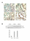
Figure 1
Figure 1
HIMF enhances VEGF expression in mouse lungs. Recombinant HIMF protein was intratracheally instilled into adult mice (200 ng/animal in 40 μl saline, n = 6 for each group). The vehicle controls were instilled with saline (40 μl/animal, n = 3). Six and twenty four hours later, the mouse lungs were collected. (A) The immunohistochemical staining results indicated that HIMF protein instillation resulted in a significant increase of VEGF production, mainly located at alveolar type II (left panels, arrows) and airway epithelial cells (right panels, arrows). Scale bars: 100 μm. (B) Western blot indicated that VEGF expression was enhanced in HIMF-instilled mouse lungs. The symbol (*) indicates a significant increase from control mouse lungs instilled with saline only (P < 0.05).
Figure 2
Figure 2
HIMF induces VEGF expression in mouse lung epithelial cell line. MLE-12 cells were treated with HIMF for various concentrations and periods as indicated. Western blots and semi-quantitative RT-PCR were performed for VEGF expression. (2A) HIMF induced VEGF protein and mRNA production in a dose-dependent manner in MLE-12 cells. (2B) Time-course study indicated that HIMF (40 nmol/L)-induced VEGF production started at 6 h, and persisted for 24 h. Triplicate experiments were performed with essentially identical results.
Figure 3
Figure 3
Generation of HIMF overexpressing cells. MLE-12 cells were transfected with HIMF cDNA or control vector. Stable cell lines, MLE-HIMF, along with their transfection control cells MLE-Zeo, were screened based on resistance to Zeocin (400 μg/ml). Western blots with cell culture medium for HIMF and protein from cell lysate for VEGF (3A) and RT-PCR with cell total RNA (3B) demonstrated that MLE-HIMF have higher HIMF protein and mRNA levels than their parent (MLE-12) and transfection (MLE-Zeo) counterparts. The VEGF levels in MLE-HIMF were also increased significantly compared with those of their controls. The symbol (*) indicates a significant increase from parent controls (P < 0.05). Triplicate experiments were performed with essentially identical results.
Figure 4
Figure 4
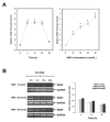
HIMF increases the transcription activities, but not mRNA stability of VEGF in MLE-12 cells. (4A) MLE-12 cells were co-transfected with pGL-VEGF and pRL-TK. Twenty-four hours later, the cells were incubated with HIMF protein as indicated. Then, cells were lysed with passive lysis buffer, and luciferase activity was measured according to the dual-luciferase assay manual. The time-course study demonstrated that HIMF-induced (40 nmol/L) VEGF transcription started at 6 h, and persisted for 24 h. After incubation with 10–80 nmol/L of HIMF, VEGF transcripts in MLE-12 were enhanced in a dose-dependent manner. (4B) MLE-12 were treated with different concentrations of HIMF and incubated with 5 μg/ml of Actinomycin D for 4, 8 and 16 h. RT-PCR indicated that HIMF did not prevent Actinomycin D-facilitated VEGF degradation in MLE-12 cells. The symbol (*) indicates a significant increase from MLE-12 controls without HIMF (P < 0.05). Triplicate experiments were performed with essentially identical results.
Figure 5
Figure 5
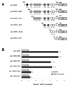
Promoter deletion assay for HIMF-induced VEGF expression in MLE-12 cells. MLE-12 cells were co-transfected with pRL-TK and each VEGF luciferase reporter construct (5A) for 24 h, then cells were incubated with HIMF protein (40 nmol/L) for another 24 h. Luciferase activity was measured and the firefly luciferase signal was normalized to the renilla luciferase signal for each individual well. (5B) Deletion of NF-κB binding site, but not HRE or binding sites for AP-1 and AP-2, completely abolished HIMF-induced VEGF promoter activity. Deletion of all cis-acting elements resulted in complete loss of induction of VEGF promoter activity. The symbol (*) indicates a significant increase from MLE-12 controls untreated with HIMF (P < 0.05). The symbol (#) indicates a significant decrease from MLE-12 transfected with pGL-VEGF and treated with HIMF (P < 0.05). Triplicate experiments were performed with essentially identical results.
Figure 6
Figure 6
HIMF activates NF-κB in MLE-12 cells. Cells were co-transfected with pNFκB-luc and pRL-TK, with or without stimulation of HIMF protein for various periods as indicated. (6A) Dual-luciferase assay indicated that MLE-HIMF had higher NF-κB activity than their control counterparts. (6B) Dual-luciferase assay indicated that HIMF protein increased the NF-κB activity in MLE-12 cells in a dose-dependent manner. The symbol (*) indicates a significant increase from MLE-12 parent controls or controls untreated with HIMF (P < 0.05). Triplicate experiments were performed with essentially identical results.
Figure 7
Figure 7
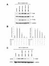
Activation of NF-κB is essential for HIMF-induced VEGF expression. Cells were co-transfected with pNFκB-luc, dominant-negative mutants of NF-κB pathway and pRL-TK, with or without stimulation of HIMF protein for various periods as indicated. (7A) Western blots indicated that HIMF (40 nmol/L) induced phosphorylation of IKK and IκBα in MLE-12 cells. (7B and 7C) Transfection of MLE-12 cells with dominant-negative mutants IKKα (K44A) and IKKβ (K44A), and super-repressor IκBα (S32A/S36A), abolished HIMF (40 nmol/L)-induced NF-κB activity and upregulation of VEGF in MLE-12 cells. The symbol (*) indicates a significant increase from MLE-12 controls untreated with HIMF (P < 0.05). The symbol (#) indicates a significant decrease from MLE-12 cells treated with HIMF only (P < 0.05). Triplicate experiments were performed with essentially identical results.
Figure 8
Figure 8
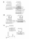
HIMF-induced NF-κB activation and upregulation of VEGF is PI-3K/Akt pathway dependent. MLE-12 cells were pretreated with signal transduction inhibitors or co-transfected with luciferase constructs and PI-3K dominant-negative mutant, then stimulated with HIMF (40 nmol/L) for various periods as indicated. (8A) HIMF strongly induced the phosphorylation of Akt at Ser473 and Thr308. The Akt phosphorylation started at 30 minutes and sustained for 360 min. HIMF also induced phosphorylation of ERK1/2 and p38 MAPK, but not JNK MAPK in MLE-12 cells. (8B) The PI-3K inhibitor LY294002 (10 μmol/L), but not SB203580 (5 μmol/L), PD098059 (5 μmol/L) or U0126 (5 μmol/L), abolished HIMF-induced Akt phosphorylation and upregulation of VEGF in MLE-12 cells. (8C) Transfection of Δp85, into MLE-12 cells abolished HIMF-induced phosphorylation of IKK and IκBα, NF-κB activation and production of VEGF. The symbol (*) indicates a significant increase from MLE-12 controls without HIMF treatment (P < 0.05). The symbol (#) indicates a significant decrease from MLE-12 cells treated with HIMF only (P < 0.05). Triplicate experiments were performed with essentially identical results.

Similar articles

Cited by

References

    1. Ferrara N. Molecular and biological properties of vascular endothelial growth factor. Journal of Molecular Medicine. 1999;77:527–543. doi: 10.1007/s001099900019. - DOI - PubMed
    1. Zeng X, Wert SE, Federici R, Peters KG, Whitsett JA. VEGF enhances pulmonary vasculogenesis and disrupts lung morphogenesis in vivo. Dev Dyn. 1998;211:215–227. doi: 10.1002/(SICI)1097-0177(199803)211:3<215::AID-AJA3>3.0.CO;2-K. - DOI - PubMed
    1. Carmeliet P, Ferreira V, Breier G, Pollefeyt S, Kieckens L, Gertsenstein M, et al. Abnormal blood vessel development and lethality in embryos lacking a single VEGF allele. Nature. 1996;380:435–439. doi: 10.1038/380435a0. - DOI - PubMed
    1. Ferrara N, Carver-Moore K, Chen H, Dowd M, Lu L, O'Shea KS, et al. Heterozygous embryonic lethality induced by targeted inactivation of the VEGF gene. Nature. 1996;380:439–442. doi: 10.1038/380439a0. - DOI - PubMed
    1. Berse B, Brown LF, Van de Water L, Dvorak HF, Senger DR. Vascular permeability factor (vascular endothelial growth factor) gene is expressed differentially in normal tissues, macrophages, and tumors. Mol Biol Cell. 1992;3:211–220. - PMC - PubMed

MeSH terms