. 2006 Mar;188(6):2073-80.
doi: 10.1128/JB.188.6.2073-2080.2006.

A complete lipopolysaccharide inner core oligosaccharide is required for resistance of Burkholderia cenocepacia to antimicrobial peptides and bacterial survival in vivo

Affiliations

A complete lipopolysaccharide inner core oligosaccharide is required for resistance of Burkholderia cenocepacia to antimicrobial peptides and bacterial survival in vivo

Slade A Loutet et al. J Bacteriol. 2006 Mar.

Abstract

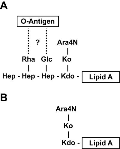
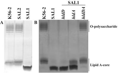
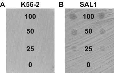
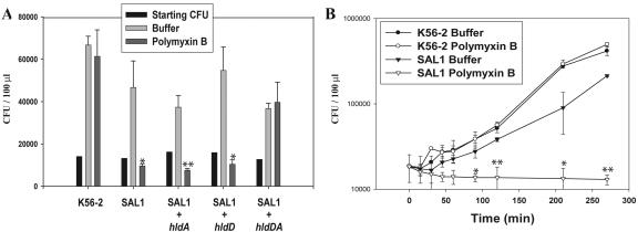
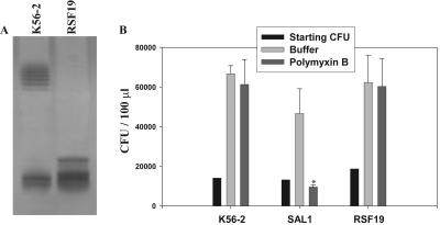
Burkholderia cenocepacia is an important opportunistic pathogen of patients with cystic fibrosis. This bacterium is inherently resistant to a wide range of antimicrobial agents, including high concentrations of antimicrobial peptides. We hypothesized that the lipopolysaccharide (LPS) of B. cenocepacia is important for both virulence and resistance to antimicrobial peptides. We identified hldA and hldD genes in B. cenocepacia strain K56-2. These two genes encode enzymes involved in the modification of heptose sugars prior to their incorporation into the LPS core oligosaccharide. We constructed a mutant, SAL1, which was defective in expression of both hldA and hldD, and by performing complementation studies we confirmed that the functions encoded by both of these B. cenocepacia genes were needed for synthesis of a complete LPS core oligosaccharide. The LPS produced by SAL1 consisted of a short lipid A-core oligosaccharide and was devoid of O antigen. SAL1 was sensitive to the antimicrobial peptides polymyxin B, melittin, and human neutrophil peptide 1. In contrast, another B. cenocepacia mutant strain that produced complete lipid A-core oligosaccharide but lacked polymeric O antigen was not sensitive to polymyxin B or melittin. As determined by the rat agar bead model of lung infection, the SAL1 mutant had a survival defect in vivo since it could not be recovered from the lungs of infected rats 14 days postinfection. Together, these data show that the B. cenocepacia LPS inner core oligosaccharide is needed for in vitro resistance to three structurally unrelated antimicrobial peptides and for in vivo survival in a rat model of chronic lung infection.


Figures

FIG. 1.
FIG. 1.
Structural diagram of full-length and heptoseless lipopolysaccharide in B. cenocepacia. The tentative structure of lipid A-core oligosaccharide was determined from the information available for B. cepacia strains GIFU 645 (28) and ATCC 25416 (19). (A) Dotted lines and the question mark indicate that the linkage with the O antigen in strain K56-2 is not known. (B) Proposed lipid A-core structure of the heptoseless LPS mutant SAL1. Kdo, deoxy-manno-octulosonic acid; Ko, glycero-talo-octulosonic acid.
FIG. 2.
FIG. 2.
Genomic organization of the hld gene cluster. The solid boxes represent internal fragments used for mutagenesis. The dotted line represents the region amplified by reverse transcription-PCR, demonstrating cotranscription of hldA and hldD.
FIG. 3.
FIG. 3.
Complementation of the E. coli LPS mutants with B. cenocepacia genes. (A) E. coli SØ874 heptokinase (hldE1) mutant FAM3 complemented with the B. cenocepacia hldA gene. (B) E. coli SØ874 epimerase (hldD) mutant SAL7 complemented with the B. cenocepacia hldD gene. − and +, absence and presence, respectively, of 0.2% rhamnose for induction of gene expression.
FIG. 4.
FIG. 4.
LPS phenotypes of SAL1 and SAL2 and complementation of SAL1. (A) Disruption of hldA results in LPS with a smaller core and no O antigen, and disruption of the downstream gene does not alter the LPS phenotype. (B) Complementation of SAL1. Both hldA and hldD are required for complementation of SAL1. Gene expression by complementing plasmids was induced with 0.2% rhamnose.
FIG. 5.
FIG. 5.
Heptoseless B. cenocepacia is sensitive to HNP-1. (A) Growth of K56-2 was not impaired at any concentration of HNP-1 tested. (B) Growth of SAL1 was inhibited at all concentrations of HNP-1 tested, as visualized by circles of growth inhibition where the plate was inoculated with HNP-1. The numbers indicate the concentrations of HNP-1 (in μg/ml) spotted in each row.
FIG. 6.
FIG. 6.
Heptoseless B. cenocepacia is sensitive to polymyxin B. (A) Number of CFU prior to challenge (Starting CFU) and 2 h after incubation in the absence (Buffer) or presence (Polymyxin B) of 100 μg/ml polymyxin B. (B) Time course analysis of growth in the absence (solid symbols) or presence (open symbols) of 100 μg/ml polymyxin B for 4.5 h. The data points and error bars indicate means and standard deviations of data from one representative experiment done in triplicate. Significant differences were determined using unpaired t tests. One asterisk indicates that the P value is <0.05 for the statistical difference between the polymyxin B and buffer treatments, and two asterisks indicate that the P value is <0.001.
FIG. 7.
FIG. 7.
Lack of O antigen does not impair resistance to polymyxin B. (A) LPS phenotypes of K56-2 and RSF19. (B) Number of CFU prior to challenge (Starting CFU) and 2 h after incubation in the absence (Buffer) or presence (Polymyxin B) of 100 μg/ml polymyxin B. The data points and error bars indicate means and standard deviations of data from one representative experiment done in triplicate. Significant differences were determined using unpaired t tests. The asterisk indicates that the P value is <0.05 for the statistical difference between the polymyxin B and buffer treatments.

References

    1. Banemann, A., H. Deppisch, and R. Gross. 1998. The lipopolysaccharide of Bordetella bronchiseptica acts as a protective shield against antimicrobial peptides. Infect. Immun. 66:5607-5612. - PMC - PubMed
    1. Bayer, M. E., J. Koplow, and H. Goldfine. 1975. Alterations in envelope structure of heptose-deficient mutants of Escherichia coli as revealed by freeze-etching. Proc. Natl. Acad. Sci. USA 72:5145-5149. - PMC - PubMed
    1. Belas, R., J. Manos, and R. Suvanasuthi. 2004. Proteus mirabilis ZapA metalloprotease degrades a broad spectrum of substrates, including antimicrobial peptides. Infect. Immun. 72:5159-5167. - PMC - PubMed
    1. Brogden, K. A. 2005. Antimicrobial peptides: pore formers or metabolic inhibitors in bacteria? Nat. Rev. Microbiol. 3:238-250. - PubMed
    1. Brötz, H., G. Bierbaum, K. Leopold, P. E. Reynolds, and H. G. Sahl. 1998. The lantibiotic mersacidin inhibits peptidoglycan synthesis by targeting lipid II. Antimicrob. Agents Chemother. 42:154-160. - PMC - PubMed

Publication types

MeSH terms

LinkOut - more resources