. 2006 Jun 1;90(11):4119-27.
doi: 10.1529/biophysj.105.078147. Epub 2006 Mar 2.

Ablation of myosin-binding protein-C accelerates force development in mouse myocardium

Affiliations

Ablation of myosin-binding protein-C accelerates force development in mouse myocardium

Julian E Stelzer et al. Biophys J. .

Abstract

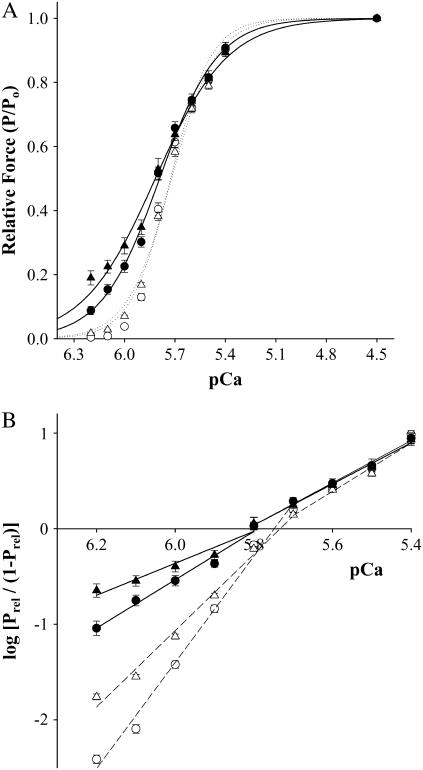
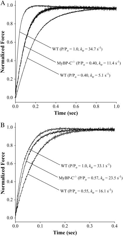
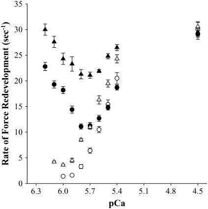
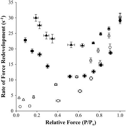
Myosin-binding protein-C (MyBP-C) is a thick filament-associated protein that binds tightly to myosin. Given that cMyBP-C may act to modulate cooperative activation of the thin filament by constraining the availability of myosin cross-bridges for binding to actin, we investigated the role of MyBP-C in the regulation of cardiac muscle contraction. We assessed the Ca(2+) sensitivity of force (pCa(50)) and the activation dependence of the rate of force redevelopment (k(tr)) in skinned myocardium isolated from wild-type (WT) and cMyBP-C null (cMyBP-C(-/-)) mice. Mechanical measurements were performed at 22 degrees C in the absence and presence of a strong-binding, nonforce-generating analog of myosin subfragment-1 (NEM-S1). In the absence of NEM-S1, maximal force and k(tr) and the pCa(50) of isometric force did not differ between WT and cMyBP-C(-/-) myocardium; however, ablation of cMyBP-C-accelerated k(tr) at each submaximal force. Treatment of WT and cMyBP-C(-/-) myocardium with 3 muM NEM-S1 elicited similar increases in pCa(50,) but the effects of NEM-S1 to increase k(tr) at submaximal forces and thereby markedly reduce the activation dependence of k(tr) occurred to a greater degree in cMyBP-C(-/-) myocardium. Together, these results support the idea that cMyBP-C normally acts to constrain the interaction between myosin and actin, which in turn limits steady-state force development and the kinetics of cross-bridge interaction.


Figures

FIGURE 1
FIGURE 1
Effect of NEM-S1 on force-pCa relationships in skinned myocardium from WT and cMyBP-C−/−. (A) All values are mean ± SE. Smooth lines were generated by fitting the mean data to a Hill equation: P/Po = [Ca2+]n/(kn + [Ca2+]n), where n is the Hill coefficient and k is the [Ca2+] required for half-maximal activation (pCa50). The relationships between Ca2+-activated force and pCa were determined in the absence (open symbols) and presence (solid symbols) of 3 μM NEM-S1. pCa50 values for WT were control (○), 5.73 ± 0.01; 3 μM NEM-S1 (•), 5.79 ± 0.01, whereas pCa50 values for cMyBP-C−/− were control (Δ), 5.72 ± 0.01; 3 μM NEM-S1 (▴), 5.82 ± 0.01. (B) Hill plot transformations of the force-pCa data were generated using the following equation: log[Prel/(1 − Prel)] = n(log[Ca2+] + k), where Prel is force as a fraction of Po, n is the Hill coefficient, and k is the [Ca2+] required for half-maximal activation (pCa50). All values are mean ± SE. Fiber characteristics are listed in Table 1.
FIGURE 2
FIGURE 2
Rate of force redevelopment in skinned myocardium from WT and cMyBP-C−/−. Differences in the rate constant of force redevelopment (ktr) were expressed relative to respective peak forces. (A) The rates of force redevelopment measured in the absence of NEM-S1 from two representative skinned myocardial preparations from either WT (left trace: pCa 4.5, P/Po = 1.0, ktr = 34.7 s−1; right trace: pCa 5.8, P/Po = 0.40, ktr = 5.1 s−1) or cMyBP-C−/− (middle trace: pCa 5.8, P/Po = 0.40, ktr = 11.4 s−1). (B) The rates of force redevelopment measured in the presence of NEM-S1 from two representative skinned myocardial preparations from either WT (left trace: pCa 4.5, P/Po = 1.0, ktr = 33.0 s−1; right trace: pCa 5.8, P/Po = 0.55, ktr = 16.1 s−1) or cMyBP-C−/− (middle trace: pCa 5.8, P/Po = 0.57, ktr = 23.5 s−1).
FIGURE 3
FIGURE 3
Effect of NEM-S1 on the Ca2+ dependence of the rate of force redevelopment. Force redevelopment after rapid release and restretch was measured as a function of pCa in skinned myocardium from WT (○,•) and cMyBP-C−/− (Δ,▴) in the absence (open symbols) and presence (solid symbols) of 3 μM NEM-S1. All values are mean ± SE. Fiber characteristics are listed in Table 2.
FIGURE 4
FIGURE 4
Effect of NEM-S1 on the activation dependence of the rate of force redevelopment. Force redevelopment after rapid release and restretch was measured as a function of relative steady-state isometric force (P/Po) in skinned myocardium from WT (○,•) and cMyBP-C−/− (Δ,▴) in the absence (open symbols) and presence (solid symbols) of 3 μM NEM-S1. All values are mean ± SE. Fiber characteristics are listed in Table 2.

References

    1. Solaro, R. J., and H. M. Rarick. 1998. Troponin and tropomyosin: proteins that switch on and tune in the activity of cardiac myofilaments. Circ. Res. 83:471–480. - PubMed
    1. Lehrer, S. S. 1994. The regulatory switch of the muscle thin filament: Ca2+ or myosin heads. J. Muscle Res. Cell Motil. 15:232–236. - PubMed
    1. Swartz, D. R., R. L. Moss, and M. L. Greaser. 1996. Calcium alone does not fully activate the thin filament for S1 binding to rigor myofibrils. Biophys. J. 71:1891–1904. - PMC - PubMed
    1. Gordon, A. M., E. Homsher, and M. Regnier. 2000. Regulation of contraction in striated muscle. Physiol. Rev. 80:853–924. - PubMed
    1. Johnson, J. D., J. H. Collins, S. P. Robertson, and J. D. Potter. 1980. A fluorescent probe study of Ca2+ binding to the Ca2+ specific sites of cardiac troponin and troponin C. J. Biol. Chem. 255:9635–9640. - PubMed

Publication types

LinkOut - more resources