. 2006 May 15;90(10):3469-84.
doi: 10.1529/biophysj.105.074377. Epub 2006 Mar 2.

Force-induced adsorption and anisotropic growth of focal adhesions

Affiliations

Force-induced adsorption and anisotropic growth of focal adhesions

Achim Besser et al. Biophys J. .

Abstract

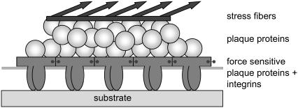
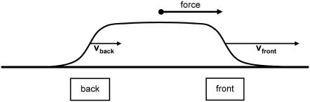
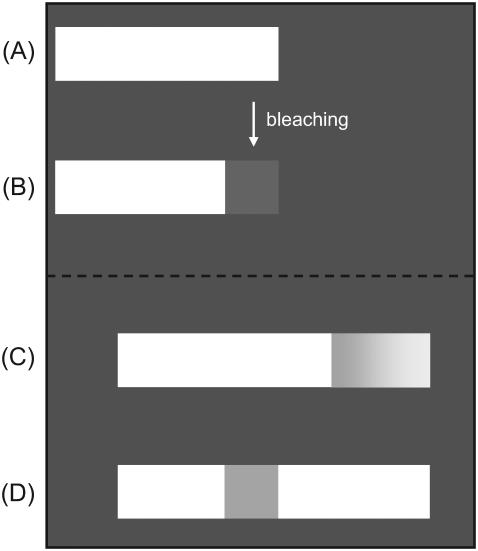
Focal adhesions are micrometer-sized protein aggregates that connect actin stress fibers to the extracellular matrix, a network of macromolecules surrounding tissue cells. The actin fibers are under tension due to actin-myosin contractility. Recent measurements have shown that as the actin force is increased, these adhesions grow in size and in the direction of the force. This is in contrast to the growth of condensed domains of surface-adsorbed molecules in which the dynamics are isotropic. We predict these force-sensitive, anisotropic dynamics of focal adhesions from a model for the adsorption of proteins from the cytoplasm to the adhesion site. Our theory couples the mechanical forces and elasticity to the adsorption dynamics via force-induced conformational changes of molecular-sized mechanosensors located in the focal adhesion. We predict the velocity of both the front and back of the adhesion as a function of the applied force. In addition, our results show that the relative motion of the front and back of the adhesion is asymmetric and in different ranges of forces, the adhesion can either shrink or grow in the direction of the force.


Figures

FIGURE 1
FIGURE 1
A schematic picture of a focal adhesion (FA) site with the actin stress fibers on top, which are connected to the plaque proteins, the so-called upper layer. The FA is grafted to the substrate by the integrin proteins. The integrin proteins together with force-sensitive plaque proteins build up the so-called lower layer.
FIGURE 2
FIGURE 2
The lower layer of integrin-protein units is modeled as a one-dimensional linear elastic chain that is anchored to the substrate. Each integrin-protein unit is represented by a particle that is connected via springs of stiffness k to its neighbors. The grafting to the surface is accounted for by a spring of stiffness kb that connects the integrin-protein unit to the substrate. The average spacing between two integrin-protein units is given by a and un is the displacement of the nth particle from its equilibrium position in the absence of force. The anchoring to the substrate by the springs of stiffness kb gives rise to a local restoring force fa,n = −kbun. Eq. 1 is a continuum representation of this discretized illustration. A derivation is given in Nicolas et al. (14).
FIGURE 3
FIGURE 3
A possible, concrete but schematic picture of the activation process of mechanosensors: Assume that the conformation of the nonactivated state of the force-sensitive proteins has a spherical shape (A), whereas the conformation of the activated state has an elongated shape (B,C). The difference in energy between the activated and nonactivated states is ΔG > 0. This energy can be provided by the external force either by compressing (B) or by stretching the sensor (C), so that the activated state is favored energetically. In-plane compression originates from elastic interaction between neighboring sensors in the lower layer, whereas stretching is due to the direct coupling of the sensor to the actin stress fibers.
FIGURE 4
FIGURE 4
The direction of the force exerted on the FA determines its front and its back edge. In the absence of artificially applied forces, the force direction is given by the actin stress fibers and generally points to the cell body. We will refer to the edge of the FA that points toward the force center (and in the ordinary case to the center of the cell) as the front edge; and the opposite boundary (generally furthest away from the center of the cell) is called the back edge. The region of FA is tracked by the plaque protein concentration profile (obtained by a cross section through the FA along the force). By calculating the change in time of plaque protein concentration at the front edge and at the back edge, experiments can separately measure the dynamics of the front and the back edges of the FA.
FIGURE 5
FIGURE 5
Numerical solution of Eq. 25 for the plaque protein concentration as a function of space and time for a constant force in the regime of a growing domain. The initial concentration profile at t = t0 is a smooth, steplike function. At early times, the front undergoes an equilibration process and the shape of the plaque protein concentration profile varies. However, after a certain time, the front begins to move in the direction of the force, in a self-similar manner that preserves the shape of the domain.
FIGURE 6
FIGURE 6
Velocity of the front and the back end of the FA as a function of the force. To obtain dimensionless units, the velocities are scaled by the factor υ0 (Eq. 36) and the force is scaled by the factor ρc, which is the critical force level at which the FA neither shrinks nor grows and Δμ0 = 0.
FIGURE 7
FIGURE 7
The overall growth velocity υtot of the FA as a function of the force. This represents the time rate of change of the overall size of the FA. The velocity is again scaled by the factor υ0 and the force by ρc.
FIGURE 8
FIGURE 8
Our model predicts different growth behavior in the different regimes of applied or actin-myosin-induced force: For small values of the force, the direct stretching of the integrin-protein units is not large enough to overcome the energy required for conformational change and the FA shrinks (R0). As the force is increased, the mechanosensors in the FA are activated; they then associate with plaque proteins from the cytosol and the loss of proteins is reduced. Because the compression term induces activation at the front edge, but deactivation by dilation at the back edge of the FA, it is the front edge that first changes (LI) its growth behavior from shrinking to a net gain of proteins (RI). For a certain force level, corresponding to a critical value of the plaque proteins' chemical potential, Δμ0 = 0, the loss at the back is exactly compensated by the aggregation of new proteins at the front (LII) and the focal adhesion neither shrinks nor grows in size. When the force is increased from this critical value, the adhesion begins to grow in size (RII). Since cells are highly dynamic structures whose growth can be rapidly changed from shrinking to growing by their contractile apparatus, cells may regulate the actin stress so that FA operates near this critical force level (LII). In the regime of very high forces the model predicts that even the back of the adhesion tends to grow (in the opposite direction of the force) due to the direct stretching effect that dominates the dilation effect (RIII). A detailed description of each force regime is given in the text.
FIGURE 9
FIGURE 9
(A) The light-colored rectangle schematically represents an FA stained with a fluorescent dye. (B) The front of the FA is bleached with a laser beam. Predicted development of the bleached region after the stimulation of the intracellular contractile apparatus or application of an external force: (C) by the model of Shemesh et al. (14); (D) by the model presented in this article.

References

    1. Geiger, B., A. Bershadsky, R. Pankov, and K. M. Yamada. 2001. Transmembrane extracellular matrix-cytoskeleton crosstalk. Nat. Rev. Mol. Cell Biol. 2:793–805. - PubMed
    1. Giancotti, F. G., and E. Ruoslahti. 1999. Integrin signaling. Science. 285:1028–1032. - PubMed
    1. Martin, P. 1997. Wound healing: aiming for perfect skin regeneration. Science. 276:75–81. - PubMed
    1. Ridley, A. J., P. M. Comoglio, and A. Hall. 1995. Regulation of scatter factor/hepatocyte growth factor responses by Ras, Rac, and Rho in MDCK cells. Mol. Cell. Biol. 15:1110–1122. - PMC - PubMed
    1. Riveline, D., E. Zamir, N. Q. Balaban, U. S. Schwarz, T. Ishizaki, S. Narumiya, Z. Kam, B. Geiger, and A. D. Bershadsky. 2001. Focal contacts as mechanosensors: externally applied local mechanical force induces growth of focal contacts by an mDia1-dependent and ROCK-independent mechanism. J. Cell Biol. 153:1175–1185. - PMC - PubMed

Publication types

LinkOut - more resources