

Functional and structural deficits in brain regions subserving face perception in schizophrenia
- PMID: 16513867
- PMCID: PMC2773688
- DOI: 10.1176/appi.ajp.163.3.455
Functional and structural deficits in brain regions subserving face perception in schizophrenia
Abstract
Objective: Schizophrenia impairs many cognitive functions, including face perception. Veridical face perception is critical for social interaction, including distinguishing friend from foe and familiar from unfamiliar faces. The main aim of this study was to determine whether patients with schizophrenia show less activation in neural networks related to face processing, compared with healthy subjects, and to investigate the relationships between this functional abnormality and anatomical abnormalities in the fusiform gyrus shown with magnetic resonance imaging (MRI).
Method: Twenty male chronic schizophrenia patients and 16 healthy comparison subjects matched with the patients for age, gender, handedness, and parental socioeconomic status underwent high-spatial-resolution MRI. Event-related potentials elicited by images of faces, cars, and hands were recorded in a separate session.
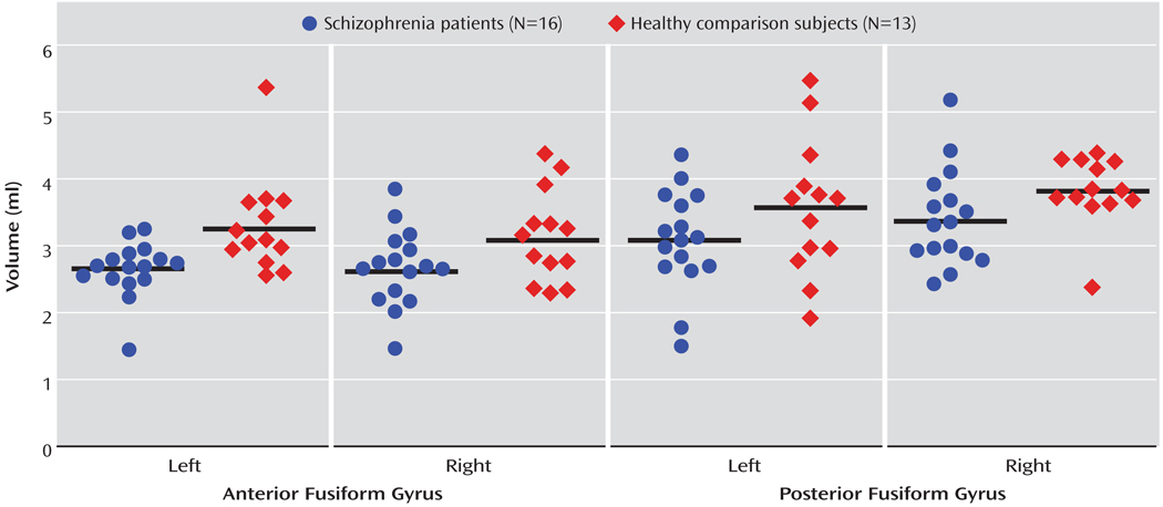
Results: Compared to the healthy subjects, the patients with schizophrenia showed bilateral N170 amplitude reduction in response to images of faces but not to images of other objects. The patients also had smaller bilateral anterior and posterior fusiform gyrus gray matter volumes, compared to the healthy subjects. In addition, right posterior fusiform gyrus volume was significantly correlated with N170 amplitude measured at the right posterior temporal electrode site in response to images of faces in the schizophrenia patients but not in the healthy comparison subjects.
Conclusions: The results provide evidence for deficits in the early stages of face perception in schizophrenia. The association of these deficits with smaller fusiform gyrus volume in patients with schizophrenia, relative to healthy subjects, suggests that the fusiform gyrus is the site of a defective anatomical substrate for face processing in schizophrenia.
Figures


Comment in
-
Understanding the glass ceiling for functional outcome in schizophrenia.Am J Psychiatry. 2006 Mar;163(3):356-8. doi: 10.1176/appi.ajp.163.3.356. Am J Psychiatry. 2006. PMID: 16513851 No abstract available.
References
-
- Gruzelier JH, Wilson L, Liddiard D, Peters E, Pusavat L. Cognitive asymmetry patterns in schizophrenia: active and withdrawn syndromes and sex differences as moderators. Schizophr Bull. 1999;25:349–362. - PubMed
-
- Penn DL, Combs D. Modification of affect perception deficits in schizophrenia. Schizophr Res. 2000;46:217–229. - PubMed
-
- Whittaker JF, Deakin JF, Tomenson B. Face processing in schizophrenia: defining the deficit. Psychol Med. 2001;31:499–507. - PubMed
-
- Damasio A, Damasio H, Van Hoesen GW. Prosopagnosia: anatomic basis and behavioral mechanisms. Neurology. 1982;32:331–341. - PubMed
Publication types
MeSH terms
Grants and funding
LinkOut - more resources
Full Text Sources
Medical
