. 2006 Mar;72(3):1891-9.
doi: 10.1128/AEM.72.3.1891-1899.2006.

Transcriptional and functional analysis of oxalyl-coenzyme A (CoA) decarboxylase and formyl-CoA transferase genes from Lactobacillus acidophilus

Affiliations

Transcriptional and functional analysis of oxalyl-coenzyme A (CoA) decarboxylase and formyl-CoA transferase genes from Lactobacillus acidophilus

M Andrea Azcarate-Peril et al. Appl Environ Microbiol. 2006 Mar.

Abstract

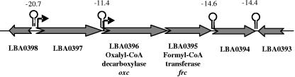
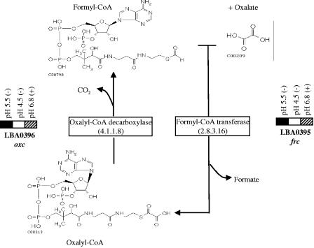
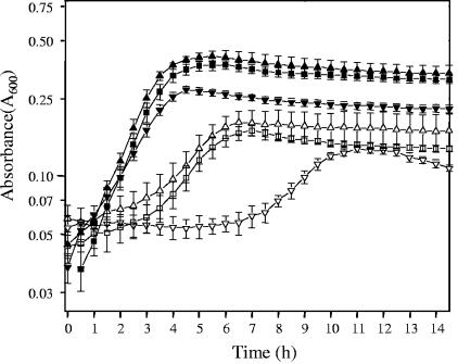
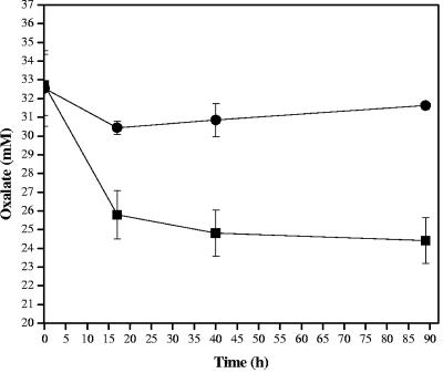
Oxalic acid is found in dietary sources (such as coffee, tea, and chocolate) or is produced by the intestinal microflora from metabolic precursors, like ascorbic acid. In the human intestine, oxalate may combine with calcium, sodium, magnesium, or potassium to form less soluble salts, which can cause pathological disorders such as hyperoxaluria, urolithiasis, and renal failure in humans. In this study, an operon containing genes homologous to a formyl coenzyme A transferase gene (frc) and an oxalyl coenzyme A decarboxylase gene (oxc) was identified in the genome of the probiotic bacterium Lactobacillus acidophilus. Physiological analysis of a mutant harboring a deleted version of the frc gene confirmed that frc expression specifically improves survival in the presence of oxalic acid at pH 3.5 compared with the survival of the wild-type strain. Moreover, the frc mutant was unable to degrade oxalate. These genes, which have not previously been described in lactobacilli, appear to be responsible for oxalate degradation in this organism. Transcriptional analysis using cDNA microarrays and reverse transcription-quantitative PCR revealed that mildly acidic conditions were a prerequisite for frc and oxc transcription. As a consequence, oxalate-dependent induction of these genes occurred only in cells first adapted to subinhibitory concentrations of oxalate and then exposed to pH 5.5. Where genome information was available, other lactic acid bacteria were screened for frc and oxc genes. With the exception of Lactobacillus gasseri and Bifidobacterium lactis, none of the other strains harbored genes for oxalate utilization.


Figures

FIG. 1.
FIG. 1.
Formyl-CoA transferase and oxalyl-CoA decarboxylase genes in L. acidophilus NCFM. Putative rho-independent terminators (lollipop symbols) and their corresponding free energies (in kcal/mol) are indicated. Potential promoter regions for ORFs LBA0396 and LBA0397 are indicated by bent arrows.
FIG. 2.
FIG. 2.
Unrooted phylogram tree of oxalyl-CoA decarboxylase sequences from diverse organisms. Proteins were aligned by CLUSTALX. Alignments were used for tree reconstruction. The organisms used were L. acidophilus NCFM, L. gasseri ATCC 3323 (GenBank accession number ZP_00046991), B. lactis (BAD11779), Bradyrhizobium japonicum USDA110 (BAC48422.1), E. coli CFT073 (NP_754791.1), Mycobacterium tuberculosis CDC1551 (NP_334536.1), O. formigenes (P40149), Mycobacterium bovis (NP_853789), Oryza sativa (BAB33274.1), Schizosaccharomyces pombe (CAA22176), Mycobacterium leprae (CAA15478), Saccharomyces cerevisiae (AAB64497), and Arabidopsis thaliana (CAC19854).
FIG. 3.
FIG. 3.
Transcriptional response of frc and oxc to pH 5.5, pH 4.5, and 1% (70 mM) ammonium oxalate (pH 6.8) in MRS broth after 30 min. The solid rectangles indicate ≥twofold-higher expression, the cross-hatched rectangles indicate a ≥twofold reduction in expression (P < 0.05), and the open rectangles indicate values of gene expression that are not statistically different from values obtained under the control conditions (L. acidophilus incubated in fresh MRS broth for 30 min). Plus and minus signs indicate that the experiment was carried out in the presence and in the absence of oxalate, respectively. The proposed metabolic pathway of oxalate decarboxylation by L. acidophilus is also shown. The structures of the compounds were obtained from the website http://www.genome.jp/kegg/kegg2.html.
FIG. 4.
FIG. 4.
Transcriptional analysis of the oxc operon in L. acidophilus cells at pH 5.5. (A) Cells were first transferred in MRS broth (pH 6.8) containing noninhibitory concentrations of ammonium oxalate (preadapted). Solid bars, frc; cross-hatched bars, oxc. Gene induction was monitored over time after cells were placed in MRS broth containing 0.5% ammonium oxalate at pH 5.5. (B) Gene induction for cells in MRS broth at pH 6.8 (nonadapted). Experiments were carried out in triplicate. The error bars indicate standard deviations.
FIG. 5.
FIG. 5.
Survival of log-phase cells of L. acidophilus NCFM and the frc mutant after challenge with MRS broth adjusted to pH 4.0, 3.5, and 3.0 with HCl, lactic acid, or oxalic acid for 2 h. The values are the averages for six separate incubations. The error bars indicate standard deviations.
FIG. 6.
FIG. 6.
Growth curves for L. acidophilus NCFM in semidefined BM containing different concentrations of ammonium oxalate. Cell growth was evaluated in BM in the presence of 0.1% glucose (▪), in the presence of glucose plus 0.1% ammonium oxalate (▴) or 0.5% ammonium oxalate (▾), in the absence of glucose (□), or in the absence of glucose plus 0.1% ammonium oxalate (▵) or 0.5% ammonium oxalate (▿). Each point represents the mean of three independent experiments. The error bars indicate standard deviations.
FIG. 7.
FIG. 7.
Oxalate-degrading activity of L. acidophilus. Strain NCFM (▪) and the frc mutant (•) were consecutively transferred in BMcit containing a noninhibitory concentration of oxalate (0.05%; 3.5 mM) and then exposed to 0.5% (32 mM) oxalate in broth. Samples were taken over time, and the oxalate concentration in the supernatants was measured. Each point represents the mean of three independent experiments. The error bars indicate standard deviations.

References

    1. Abe, K., Z.-S. Ruan, and P. C. Maloney. 1996. Cloning, sequencing, and expression in Escherichia coli of OxlT, the oxalate:formate exchange protein of Oxalobacter formigenes. J. Biol. Chem. 271:6789-6793. - PubMed
    1. Altermann, E., W. M. Russell, M. A. Azcarate-Peril, R. Barrangou, B. L. Buck, O. McAuliffe, N. Souther, A. Dobson, T. Duong, M. Callanan, S. Lick, A. Hamrick, R. Cano, and T. R. Klaenhammer. 2005. Complete genome sequence of the probiotic lactic acid bacterium Lactobacillus acidophilus NCFM. Proc. Natl. Acad. Sci. USA 102:3906-3912. - PMC - PubMed
    1. Andresson, H., S. Filipsson, and L. Hulten. 1978. Urinary oxalate excretion related to ileocolic surgery in patients with Crohn's disease. Scand. J. Gastroenterol. 13:465-469. - PubMed
    1. Axelsen, K. B., and M. G. Palmgren. 1998. Evolution of substrate specificities in the P-type ATPase superfamily. J. Mol. Evol. 46:84-101. - PubMed
    1. Azcarate-Peril, M. A., O. McAuliffe, E. Altermann, S. Lick, W. M. Russell, R. Cano, and T. R. Klaenhammer. 2005. Microarray analysis of a two-component regulatory system involved in acid resistance and proteolytic activity in Lactobacillus acidophilus. Appl. Environ. Microbiol. 71:5794-5804. - PMC - PubMed

Publication types

MeSH terms